
Count Cells without Cell Staining
Perform cell proliferation, cytotoxicity, and other assays without nuclear stains—and save up to 70 mins on your workflow
Imaging cell-based assays typically requires the use of fluorescent probes that can be toxic to living cells or may only function in fixed cells. A label-free method for analyzing cell counts and cell confluence enables researchers to quantitatively monitor cell proliferation and health without time-consuming workflows that may disrupt cell viability. Perform cell proliferation, cytotoxicity, and other assays without nuclear stains like DAPI, which intercalates with DNA, or live cell dyes that are toxic to cells in the long term. Here, we describe the analysis of a variety of commonly used cell types including NIH3T3, THP-1, HepG2, plus many more. without the use of cell staining.
Counting cells without nuclear stains
Imaging cell-based assays typically requires the use of fluorescent probes that can be toxic to living cells or may only function in fixed cells. A label-free method for analyzing cell counts and cell confluence enables you to quantitatively monitor cell proliferation and health without time-consuming workflows that may disrupt cell viability. Our multi-mode microplate reader, imaging cytometer and unique, StainFreeTM Cell Detection Technology allows you to perform cell proliferation, cytotoxicity, and other assays without nuclear stains like DAPI, which intercalates with DNA, or live cell dyes that are actually toxic to cells in the long term.
The StainFree cell detection algorithm eliminates cell staining for cell counting and confluence measurements using proprietary transmitted light (TL) analysis technology. Here, we describe the analysis of a variety of commonly used cell types using our label-free method.
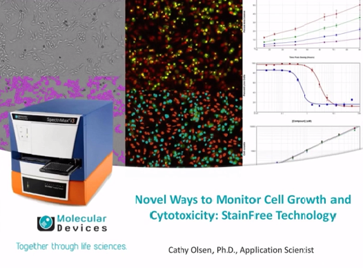
Monitor cell growth and cytotoxicity without labels using StainFree technology
In this video, Novel Ways to Monitor Cell Growth and Cytotoxicity, learn how our StainFree technology is used in applications including:
- Cell counting
- Monitoring cell growth over time
- Cytotoxicity assay
- Expression analysis
- Viral plaque assays
And, learn more about our SpectraMax i3x platform.
About StainFree Cell Detection Technology
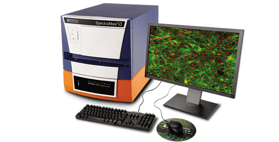
Counting cells and phenotypic characterization are important subsets of cytometry used in various applications within life science research. Our SpectraMax® i3x Multi-Mode Microplate Reader with the MiniMaxTM 300 Imaging Cytometer enables cellular imaging assays in a small footprint and eliminates cell staining for cell counting and confluence measurements with StainFree Cell Detection Technology. This field-upgradeable option with one-of-a kind brightfield cell segmentation, green and red fluorescent channel detection, and a simple workflow provides you with cellular analysis capability without the need for a complex imaging system.
Comparison of cell analysis workflows
The StainFree workflow can save up to 70 minutes compared to fixation and staining with DAPI. Moreover, cells analyzed using StainFree technology remain fully viable and can be used in additional assays.

Count cells with the MiniMax cytometer.
Cells stained with Live Red Dye were imaged in the red fluorescent (left) or transmitted light (middle) channel. StainFree cell counts (right, purple masks) correlate to cell counts based on red nuclear staining, as shown in the graph. The r2 values for both curves were 0.99.
