
Cell Counter
At The Cell Counter : HeLa Cells
HeLa cells were the first cell lines to be grown in the lab and are still used in countless biomedical research projects today. They were derived from cervical cancer cells taken in 1951 from Henrietta Lacks, a patient who later died from the disease. Rebecca Skloot’s bestselling book “The Immortal Life of Henrietta Lacks” tells the fascinating story of her life and legacy.
View StainFree Cell Detection Webinar
Download StainFree Cell Detection Application Note
Download eBook: Count Cells Like a Pro
Figure 1. StainFree analysis compared to fluorescence cell counting
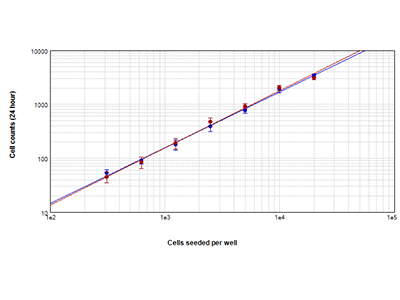
HeLa cells counted using StainFree™ Cell Detection Technology (blue circles) and red nuclear stain (red circles). Counts obtained from both methods agree closely, demonstrating that StainFree technology gives accurate cell counts while eliminating the need for fluorescent dyes (r > 0.99 for each plot).
Figure 2. Growth curves obtained using StainFree covered area analysis
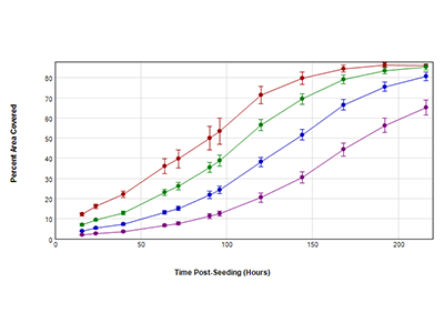
HeLa cells were seeded at 4 initial densities in a 96-well plate: 250 (purple), 500 (blue), 1000 (green), and 2000 (red) cells per well. Cells were imaged with the transmitted light (TL) channel for 9 days. Percent area covered by cells at each time point was determned with StainFree analysis.
Figure 3. Cytotoxicity measured using StainFree technology
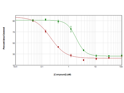
HeLa cells were seeded at 1000 cells per well and allowed to grow overnight. Then they were treated with anisomycin (red circles) or trichostatin (green circles) for 72 hours. Cytotoxicity was measured by calculating the percent area covered using StainFree analysis. IC50 curves were plotted with SoftMax Pro Software.
Figure 4. Images of HeLa cells treated with toxic compounds
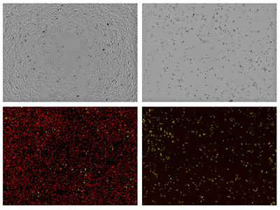
Top row: HeLa cells were treated with low (left) or high (right) anisomycin and images with the transmitted light (TL) channel. Bottom row: HeLa cells were treated with low (left) or high (right) concentrations of staurosporine and assayed for cytotoxicity with the EarlyTox Cell Integrity Kit. Live cells are labeled red and dead cells are labeled both red and green.
Tip 1:
To count HeLa cells without staining, use either the predefined setting “CellsA” in SoftMax Pro Software or create a new setting using the drawing tools in the software. The analysis method you use will depend on your cells’ morphology and growth conditions. Try the predefined setting first and see how that looks.
Tip 2:
Getting growth curves like the ones show here is simplified with the Field Analysis feature in SoftMax Pro Software. Field Analysis calculates the percent area covered by cells (confluence) in your images. Calculating cell confluence can also be helpful for assay development. For example, you can assay your cells at several different densities and determine which one yields the best assay results.
Tip 3:
Monitoring cytotoxicity over time is easy. Simply analyze the percent area covered by cells at desired time points, and plot the results in SoftMax Pro Software. There’s no need to stain or harvest your cells.
HeLa Cells Analysis Toolkit
- SpectraMax® i3 Multi-Mode Microplate Detection Platform
- SpectraMax® MiniMax™ 300 Imaging Cytometer
- SoftMax® Pro Software
- EarlyTox™ Cell Integrity Kit
Instrument Settings
Analysis type: Discrete Object Analysis
Wavelength for finding objects: TL
About StainFree Cell Detection Technology
Imaging cell-based assays typically requires the use of fluorescent probes that can be toxic to living cells or may only function in fixed cells. A label-free method for analyzing cell counts and cell confluence enables researchers to quantitatively monitor cell proliferation and health without time-consuming workflows that may disrupt cell viability.
The SpectraMax i3 Multi-Mode Microplate Platform with MiniMax 300 Imaging Cytometer uses unique, patent-pending StainFree Cell Detection Technology that allows you to perform cell proliferation, cytotoxicity, and other assays without nuclear stains like DAPI, which intercalates with DNA, or live cell dyes that are actually toxic to cells in the long term.