
Cell Counter
At the Cell Counter: THP-1 Cells
Derived from the blood of an acute monocytic leukemia patient over 40 years ago, the human monocytic cell line THP-1 is widely used to study the function of monocytes and macrophages, as well as in leukemia research. Normally THP-1 cells grow in suspension, but they can be induced to differentiate into adherent macrophage-like cells, which express IL-8 and other markers, and can thus serve as a model system for differentiation.
View StainFree Cell Detection Webinar
Download StainFree Cell Detection Application Note
Download eBook: Count Cells Like a Pro
Figure 1: StainFree and fluorescent nuclear counts of undifferentiated THP-1 cells
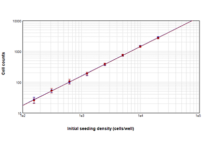
THP-1 cells seeded at densities ranging from 156 to 20,000 cells per well were counted the day after seeding using StainFree technology (blue circles), or their nuclei were counted after staining with EarlyTox Live Red Dye (red circles). Cell counts obtained with both methods differed by 5% or less at all cell densities.
Figure 2: Undifferentiated THP-1 cells imaged on the MiniMax cytometer
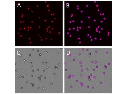
A, THP-1 cells stained with EarlyTox Live Red Dye. B, Purple object masks showing cells identified by SoftMax Pro Software in the red fluorescent imaging channel. C, Cells imaged in the TL channel. D, Cells identified by StainFree analysis in the TL channel.
Figure 3: THP-1 differentiation time course
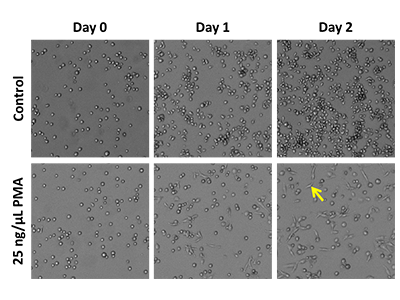
THP-1 cells were seeded at 20,000 cells per well in a 96-well black-wall, clear-bottom plate. They were treated with 25 ng/µL PMA or with DMSO control. Cells were imaged on Day 0, Day 1 post-addition, and Day 2 post-addition. The yellow arrow indicates a differentiated cell with adherent, flattened morphology.
Figure 4: THP-1 cell proliferation
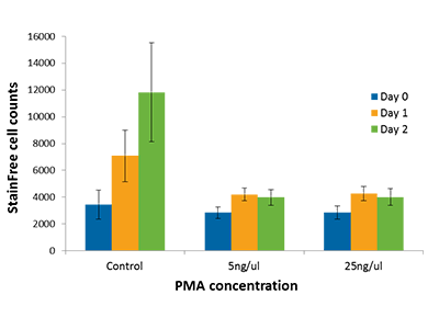
Cell proliferation was measured in PMA-treated or DMSO-treated control cells using StainFree technology. PMA treatment halts cell proliferation, but control cells continue to divide.
Figure 5: IL-8 ELISA
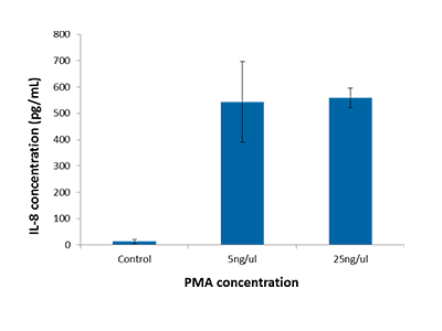
IL-8 concentration measured via ELISA in PMA- and DMSO-treated THP-1 cells. Cells were seeded at 24,000 cells per well, and supernatants were collected for assay 48 hours after treatment was initiated. The absorbance-based IL-8 ELISA was detected using a SpectraMax i3 microplate reader.
Tip :
For suspension cells, such as undifferentiated THP-1 cells, you can use a lower exposure time to minimize blurring caused by the floating motion of the cells. The SpectraMax MiniMax 300 Imaging Cytometer is able to switch wavelengths very rapidly, minimizing the amount of cell movement that can occur between images and leading to better overlay of images taken in different channels. The ‘CellsC’ predefined TL image analysis setting works well for suspension cells with rounded morphology, while ‘CellsA’ more accurately identifies the differentiated, more flattened cells.
THP-1 Cells Analysis Toolkit
- SpectraMax ® i3 Multi-Mode Microplate Detection Platform
- SpectraMax ® MiniMax™ 300 Imaging Cytometer
- SoftMax ® Pro Software
Instrument Settings for imaging and cell counting
Analysis Type: Discrete Object Analysis
Wavelength for Finding Objects: TL or 713
About StainFree Cell Detection Technology
Imaging cell-based assays typically requires the use of fluorescent probes that can be toxic to living cells or may only function in fixed cells. A label-free method for analyzing cell counts and cell confluence enables researchers to quantitatively monitor cell proliferation and health without time-consuming workflows that may disrupt cell viability.
The SpectraMax i3 Multi-Mode Microplate Platform with MiniMax 300 Imaging Cytometer uses unique, patent-pending StainFree Cell Detection Technology that allows you to perform cell proliferation, cytotoxicity, and other assays without nuclear stains like DAPI, which intercalates with DNA, or live cell dyes that are actually toxic to cells in the long term.