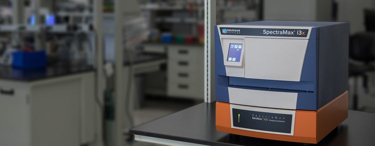

SpectraMax i3x Multi-Mode Microplate Reader
Western blot, cell counting, and injectors in one configurable microplate reader
Unlimited breadth of user-installable application modules expands research capabilities
The SpectraMax® i3x Multi-Mode Microplate Reader can perform a wide range of essential cell-based and biochemical assays using the included absorbance, fluorescence, and luminescence detection modes. With the SpectraMax® MiniMax™ 300 Imaging Cytometer, industry-leading SoftMax® Pro Software, and user-configurable detection modules, this unique reader enables users to add optional detection modules at any time to expand the reader’s capabilities and explore cellular pathways and protein expression on one microplate reader. This includes AlphaScreen/AlphaLisa, time-resolved fluorescence, TR-FRET/HTRF, fluorescence polarization, dual injectors, and western blot detection. Transmitted light and fluorescence channels offer the option to image stain-free or use cellular dyes to get a better view of cytotoxicity, cell proliferation, and phenotypic changes.

Capture flash assays with ease
The SpectraMax i3x Injector Cartridge with SmartInject® Technology expands your research capabilities to include flash-based luminescence applications including dual luciferase.

Achieve unparalleled sensitivity
The super-cooled PMT reduces background noise, providing greater sensitivity and an expanded dynamic range.

Increase fluorescence performance
An innovative combination of xenon flash lamp and LEDs provides unmatched signal strength and superior sensitivity with Spectral Fusion™ Illumination.
Features

User-configurable applications
User-installable detection modules expand the reader’s detection capabilities to include AlphaScreen, time-resolved fluorescence, TR-FRET (including HTRF), fast kinetics with injectors, western blot detection, and more.

No-stain cell analysis
The StainFree™ Cell Detection Algorithm for brightfield cell segmentation enables cell counting and confluency measurement without destructive stains, simplifying the cell analysis workflow.

Multi-channel functionality
Two fluorescence detection channels on the MiniMax 300 cytometer enable the analysis of cell viability or cell toxicity assays, including viability assays and transfection efficiency.

Cellular pathway analysis
Cell confluence and cell viability imaging, western blot analysis, and quantitation of nucleic acids and protein, can be captured on one reader.

Spectral Optimization Wizard
This built-in technology enables users to optimize their assay in a single-step by quickly identifying the best excitation and emission wavelengths.

Auto Intensity Adjustment
Our patented technology acquires data over a wide dynamic range without the need to re-read the plate due to saturation effects. PMT gain adjustment is bypassed with the use of high-power LEDs as part of the Spectral Fusion Illumination.
Add Cellular Imaging to your Plate Reader Workflow
Observe the viability of your cells with the field-upgradeable cellular imaging option for the SpectraMax i3x Multi-mode Detection Platform.
The field-upgradeable SpectraMax® MiniMax™ 300 Imaging Cytometer enables quick imaging and analysis of cells and gives you a front-row view of phenotypic changes that accompany cytotoxicity, cell proliferation, and protein expression. Now with StainFree™ Cell counting Technology, additional brightfield analysis features, and fluorescent green and red detection channels.

Latest Resources
Featured Applications
Customer Breakthrough
Applications of SpectraMax i3x Multi-Mode Microplate Reader
Specifications & Options of SpectraMax i3x Multi-Mode Microplate Reader
*Using lowest settings and speed read when available.
Download Brochure

Resources of SpectraMax i3x Multi-Mode Microplate Reader
SpectraMax® i3x Multi-Mode Microplate Reader
SpectraMax® i3x Multi-Mode Microplate Reader System
Fluorescence Polarization Detection Cartridge
(Fluorescein)
Luminescence Detection Cartridge
(Glow for 96-well plates)
Luminescence Detection Cartridge
(Glow 96- and 384-well plates)
Luminescence Detection Cartridge
(Glow 96-, 384-, and 1536-well plates)
Dual Glow Luminescence Detection Cartridge
(BRET2)
Fluorescence Intensity Detection Cartridge
(Courmarin-Fluorescein)