
Cell Counter
At the Cell Counter: U937 Cells
U937 cells were isolated from a histiocytic lymphoma of a 37-year-old male patient in 1974. Because they are one of just a few cell lines available that express many monocytic characteristics, they are often used to study the behavior and differentiation of monocytes. U937 cells undergo apoptosis when treated with granulocyte-macrophage colony-stimulating factor (GM-CSF; Okuma et al., 2000), making them a useful model for studies of apoptotic cell signaling.
View StainFree Cell Detection Webinar
Download StainFree Cell Detection Application Note
Download eBook: Count Cells Like a Pro
Figure 1 : Images obtained using SpectraMax MiniMax cytometer
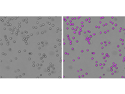
U937 cells were identified using the predefined analysis setting ‘CellsC’ in SoftMax Pro Software. Left, transmitted light image; right, StainFree analysis with purple masks indicating cells identified by the software.
Figure 2: StainFree cell counts vs. fluorescent cell counts
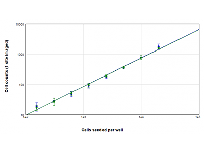
U937 cells were seeded at densities ranging from 156 to 20,000 cells per well, and nuclei were stained using EarlyTox™ Live Green dye. Images were acquired from 1 site per well. Cells in each image were then counted by using either the StainFree method (blue plot) or by counting green-fluorescent nuclei (green plot).
Figure 3: EarlyTox Live/Dead Assay
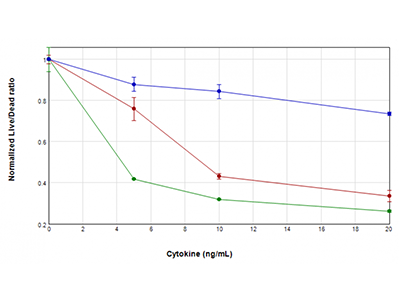
U937 cells were treated with concentrations of GM-CSF (blue plot), TNFα (red plot), or TNFα plus GM-CSF (green plot) ranging from 0 ng/mL to 20 ng/mL, for 48 hours. They were then assayed for viability using the EarlyTox™ Live/Dead Assay Kit. Fluorescence was detected using the SpectraMax i3x Multi-Mode Microplate Reader. The ratio of live/dead fluorescent signal was plotted vs. cytokine concentration, revealing a loss of cell viability in treated cells that is particularly severe when TNFα and GM-CSF are combined.
Figure 4: EarlyTox Caspase-3/7 R110 Assay
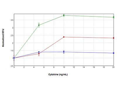
U937 cells were treated with concentrations of TNFα plus GM-CSF (green plot), TNFα (red plot), or GM-CSF only (blue plot) ranging from 0 ng/mL to 20 ng/mL, for 48 hours. They were then assayed for caspase-3/7 activity using the EarlyTox™ Caspase-3/7 R110 Assay. Fluorescent signal indicative of caspase activity was detected using the SpectraMax i3x reader. The highest caspase activity was observed when cells were treated with both TNFα and GM-CSF, suggesting a synergistic activation of caspase.
Figure 5: EarlyTox Caspase-3/7 NucView 488 Assay
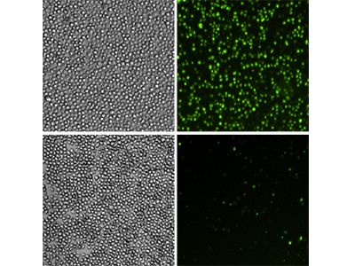
U937 cells were treated with 10 ng/mL of TNFα and GM-CSF (top row) or left untreated (bottom row) for 48 hours. They were then assayed using the EarlyTox™ Caspase-3/7 NucView™ 488 Assay . Green fluorescent staining identified the cells with active caspase-3/7. Left panels, transmitted light images of cells; right panels, green fluorescent images revealing cells with active caspase-3/7.
Tip:
U937 cells grow in suspension and have a round shape that makes them easy to count. For StainFree counting, simply use the predefined analysis setting ‘CellsC’ in the dropdown menu in SoftMax Pro Software to analyze cell images with a single click.
U937 Cells Analysis Toolkit
- SpectraMax ® i3 Multi-Mode Microplate Detection Platform
- SpectraMax ® MiniMax™ 300 Imaging Cytometer
- SoftMax ® Pro Software
Transmitted light (TL)
541 nm (green fluorescence)
TL exposure: 7 ms
TL Focus adjustment: -5 µm
541 exposure: 1 ms
541 Focus adjustment: 40 µm
Analysis type: Discrete Object Analysis
Wavelength for finding objects: TL (StainFree) or 541 (green nuclei)
TL: CellsC (predefined)
541: Set Size and Intensity: Size = 10-30 µm, Intensity Above Background = 150
About StainFree Cell Detection Technology
Imaging cell-based assays typically requires the use of fluorescent probes that can be toxic to living cells or may only function in fixed cells. A label-free method for analyzing cell counts and cell confluence enables researchers to quantitatively monitor cell proliferation and health without time-consuming workflows that may disrupt cell viability.
The SpectraMax i3/i3x Multi-Mode Microplate Platform with MiniMax 300 Imaging Cytometer uses unique, patent-pending StainFree Cell Detection Technology that allows you to perform cell proliferation, cytotoxicity, and other assays without nuclear stains like DAPI, which intercalates with DNA, or live cell dyes that are actually toxic to cells in the long term.