Stem cell science insights and breakthroughs presented at #ISSCR2021
If you didn't get a chance to visit us at our poster sessions during ISSCR 2021, don’t fret. We've gathered all our sessions right here for you.
The ISSCR Annual Meeting brought together nearly 4,000 leaders from across the globe to discuss new technologies, share research insights, and explore the newest breakthroughs in stem cell science and regenerative medicine. With in-depth scientific programs and networking opportunities, visitors gained inspiration and insights to advance their research.
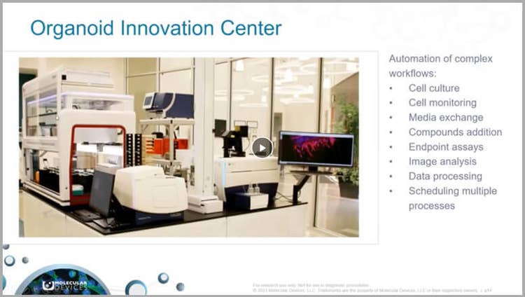
This year, we were honored to be a part of the Innovation Showcase with our session: Automated Culture and High-Content Imaging of 3D Lung and Cardiac Organoids for In Vitro Assessment of Compound Effects. Dr. Oksana Sirenko shared the why and how of automated complex 3D biological assay workflows and explained the ways new advances in high-content imaging technology are revealing significant data. This data that can help scale up organoid assays for screening and push the boundaries of your research.

Watch our on-demand virtual poster sessions
A staple of the ISSCR Annual Meeting, poster sessions allow invited guests to engage one-on-one with researchers. Here, we offer a virtual experience to view our presentations at your leisure and encourage you to contact us anytime to discuss your research challenges.
Register once to access all five of this year's poster sessions and listen to the on-demand webinars.
- Poster 1 - Innovation Showcase: Automated culture and high-content imaging of 3D lung and cardiac organoids for in vitro assessment of compound effects
- Poster 2 - Monitoring organoid development and characterization of calcium oscillation activities in iPSC-derived 3D cerebral organoids
- Poster 3 - Deep learning-based image analysis for label-free live monitoring of iPSC 3D organoid cultures
- Poster 4 - Organoids for disease modeling and in vitro drug screening: Automated culture monitoring, imaging, and analysis of complex biological systems
- Poster 5 - Assessment of compound-induced pro-arrhythmic effects in human ipsc-derived cardiomyocytes
View on-demand poster sessions
Poster 1) Innovation Showcase: Automated culture and high-content imaging of 3D lung and cardiac organoids for in vitro assessment of compound effects
Simultaneously, advances in high-content imaging technology are helping to reveal significant information from these complex biological systems, inviting scientists to push the boundaries of their research. In this webinar, we describe an integrated workflow that allows us to automate processes of cell culture, imaging, and cell maintenance to monitor the development of stem cells and organoids, as well as characterize the complex responses to test compounds. The method was applied for automation of three complex workflows: iPSC maintenance, lung toxicity assay using 3D lung organoids, and monitoring compound effects on functional activity in 3D cardiac cultures.
Watch now to view our Innovation Showcase presentation and more...
Poster 2) Monitoring organoid development and characterization of calcium oscillation activities in iPSC-derived 3D cerebral organoids
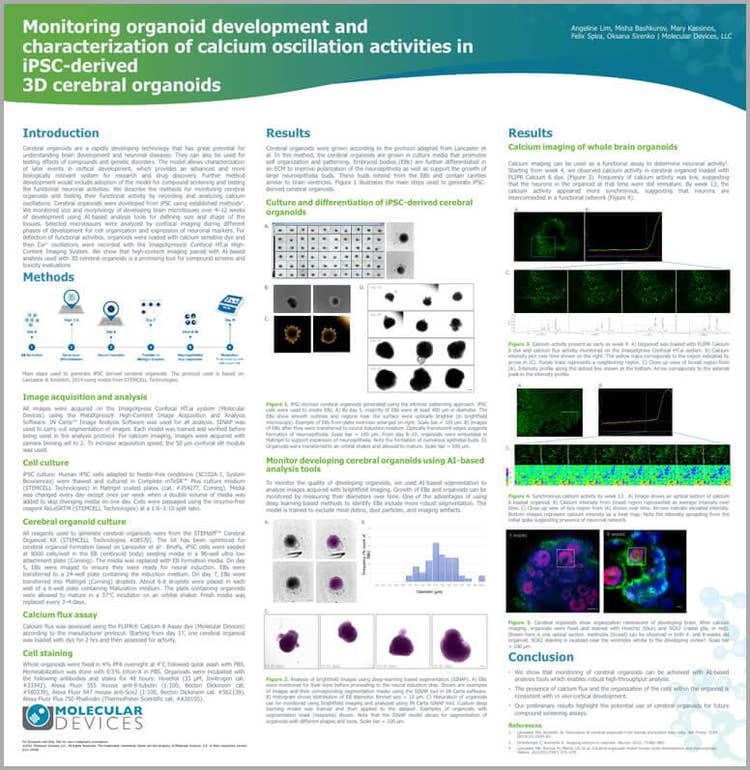
Cerebral organoids are a rapidly developing technology that has great potential for understanding brain development and neuronal diseases. They can also be used for testing effects of compounds and genetic disorders. The model allows characterization of later events in cortical development, which provides an advanced and more biologically relevant system for research and drug discovery.
Further method development would include adoption of the model for compound screening and testing the functional neuronal activities. We describe the methods for monitoring cerebral organoids and testing their functional activity by recording and analyzing calcium oscillations.
Cerebral organoids were developed from iPSC using established methods. We monitored size and morphology of developing brain microtissues over 4-12 weeks of development using AI-based analysis tools for defining size and shape of the tissues. Selected microtissues were analyzed by confocal imaging during different phases of development for cell organization and expression of neuronal markers. For detection of functional activities, organoids were loaded with calcium sensitive dye and then Ca2+ oscillations were recorded with the ImageXpress® Confocal HT.ai High-Content Imaging System. We show that high-content imaging paired with AI-based analysis used with 3D cerebral organoids is a promising tool for compound screens and toxicity evaluations.
Watch now to learn about monitoring 3D cerebral organoids and more...
Poster 3) Deep learning-based image analysis for label-free live monitoring of iPSC 3D organoid cultures
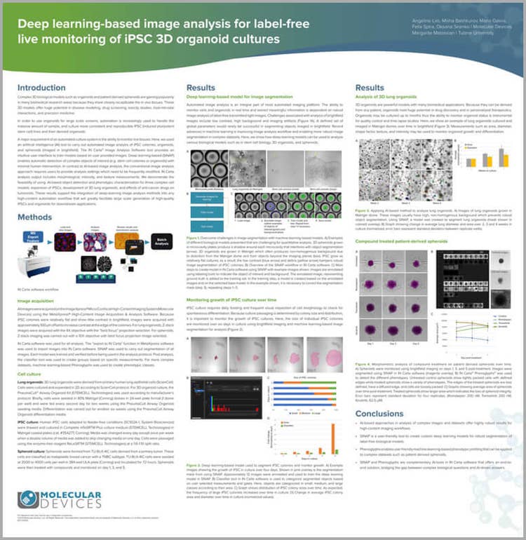
Complex 3D biological models such as organoids and patient-derived spheroids are gaining popularity in many biomedical research areas because they more closely recapitulate the in vivo tissues. These 3D models offer huge potential in disease modeling, drug screening, toxicity studies, host-microbe interactions, and precision medicine.
In order to use organoids for large scale screens, automation is increasingly used to handle the massive amount of sample, and culture more consistent and reproducible iPSC (induced pluripotent stem cell) lines and their derived organoids.
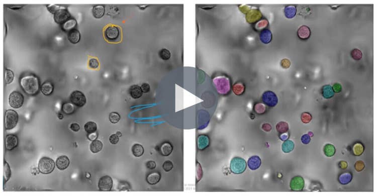
A major requirement of an automated culture system is the ability to monitor live tissues. Here, we used an artificial intelligence (AI) tool to carry out automated image analysis of iPSC colonies, organoids, and spheroids (imaged in brightfield). The IN Carta™ Image Analysis Software tool provides an intuitive user interface to train models based on user provided images. Deep learning-based (SINAP) enables automatic detection of complex objects of interest (e.g. stem cell colonies or organoids) with minimal human intervention. In contrast to AI-based image analysis, the conventional image analysis approach requires users to provide analysis settings which need to be frequently modified. IN Carta analysis output includes morphological, intensity, and texture measurements. We demonstrate the feasibility of using AI-based object detection and phenotypic characterization for three complex cell models: expansion of iPSCs, development of 3D lung organoids, and effects of anti-cancer drugs on tumoroids. These results support the integration of deep-learning image analysis methods into any high-content automation workflow that will greatly facilitate large scale generation of high-quality iPSCs and organoids for downstream applications.
Watch now to learn about AI-based image analysis and more...
Poster 4) Organoids for disease modeling and in vitro drug screening: Automated culture monitoring, imaging, and analysis of complex biological systems
We describe an automated integrated system that allows automated monitoring, maintenance, and characterization of growth and differentiation of stem cells and organoids, as well as testing the effects of various compounds. The automated integrated system included the ImageXpress Confocal HT.ai system, an automated incubator, a Biomek liquid handler, and a robotic device. We demonstrated methods for successful automation of three complex workflows: iPSC culture, 3D lung organoids, and intestinal organoids. The workflow demonstrates the usefulness of automation and advanced high-content imaging for increased throughput and information in organoid assays, critical for compound screening.
Watch now to learn about automated integrated system and more...
Poster 5) Assessment of compound-induced pro-arrhythmic effects in human ipsc-derived cardiomyocytes
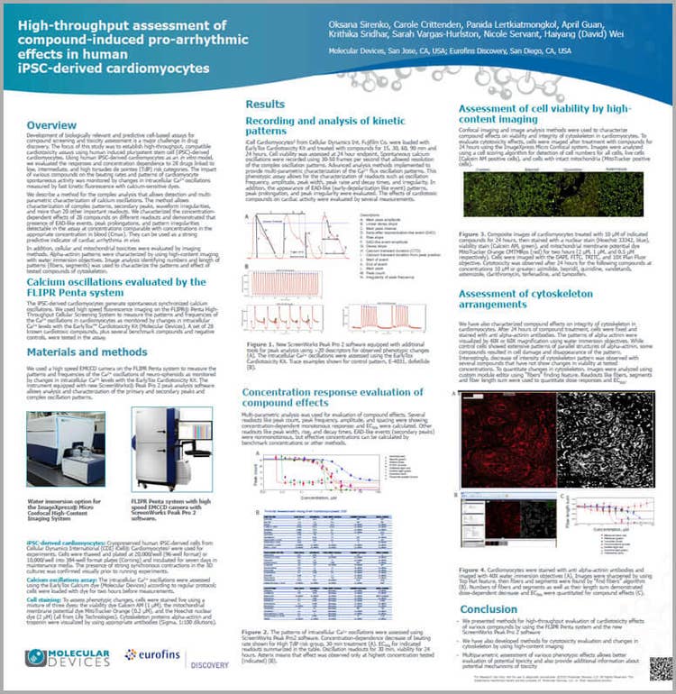
Development of biologically relevant and predictive cell-based assays for compound screening and toxicity assessment is a major challenge in drug discovery. The focus of this study was to establish high-throughput compatible cardiotoxicity assays using human induced pluripotent stem cell (iPSC)-derived cardiomyocytes.
Using human iPSC-derived cardiomyocytes as an in vitro model, we evaluated the responses and concentration dependence to 28 drugs linked to low, intermediate, and high torsades de pointes (TdP) risk categories. The impact of various compounds on the beating rates and patterns of cardiomyocyte spontaneous activity was monitored by changes in intracellular Ca2+ oscillations measured by fast kinetic fluorescence with calcium-sensitive dyes on the FLIPR® Penta High-Throughput Cellular Screening System.
We describe a method for the complex analysis of calcium oscillations that allows detection and multi-parametric characterization of oscillation peaks and patterns. In addition to detection of oscillation rates, peak width and amplitude, the method allows characterization of complex patterns, secondary peaks, waveform irregularities, and more than 20 other important readouts. Compound-induced pro-arrhythmic effects such as Early After-Depolarization (EADs)-like events or peak prolongations can be easily identified and flagged.
In addition, cellular and mitochondrial toxicity were assessed in the follow-up assay by high-content imaging. We characterized the concentration-dependent effects of 28 compounds on different readouts and demonstrated that presence of EAD-like events, peak prolongations, and pattern irregularities detectable in the assay at concentrations comparable with concentrations in the appropriate concentration in blood (Cmax), can be used as a strong predictive indicator of cardiac arrhythmia in vivo.
The results demonstrate the utility of the method for high-throughput screening and detection of drug-induced pro-arrhythmic effects in vitro.
Watch now now to learn about cardiotoxicity using iPSC-derived cardiomyocytes and more...
Explore the solutions for complex stem cell data analysis and visualization
Whether you’re just starting to explore the benefits of 3D tissue models and imaging or are leveraging advanced 3D workflows, we can offer solutions for the complexities that come with organoid and stem cell data analysis, automate your 3D culture workflows to quantify and visualize 3D structures, and help you reduce lab costs and push the boundaries of your research.