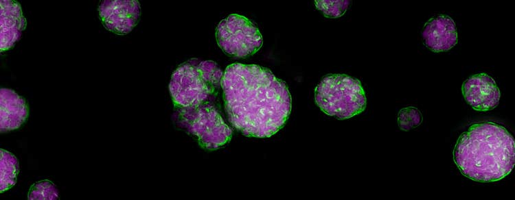
Spheroids
Application of spheroids for cancer research and drug screening
What are spheroids?
Spheroids are three-dimensional (3D) cell aggregates that can mimic tissues and microtumors. In recent years, there has been significant progress in development of in vitro aggregates of tumor cells for use as models for in vivo tissue environments. When seeded into a well of a low-attachment round bottom microplate, these aggregates will form a discrete spheroid.
Spheroids are believed to mimic tumor behavior more effectively than regular two dimensional (2D) cell cultures because, much like tumors, they contain both surface-exposed and deeply buried cells, proliferating and non-proliferating cells, and a hypoxic center with a well-oxygenated outer layer of cells. Such 3D spheroid models are being successfully used in screening environments for better assessment of drug safety and identifying potential cancer therapeutics.
The application of spheroids for cancer research and drug screening
One of the most significant objectives in cancer research is to understand tumor cell formation. Compared to 2D cell cultures, spheroids mimic solid tumors much more accurately. They help display the physiological changes that differentiate tumor cells from healthy cells. Multicellular tumor spheroid models give deeper insights into the tumor microenvironment, allowing researchers to visualize cell-to-cell interactions, how tumor cells absorb nutrients, and how they proliferate.
Spheroids are also ideal for preclinical drug screening and validation when monitoring cellular responses to therapeutic interventions. Specifically, they can reveal the tumor infiltration capacity (or incapacity) of drugs, as well as their inhibitory effects on metastasis.
Stem cell research is another significant application of spheroids, especially during the cultivation of embryonic (ESC) and neural stem cells (NSC). Recently, researchers cultured mesenchymal stem cells (MSCs) in spheroids and proved their regenerative and anti-inflammatory effects, which depicts potential for regenerative medicine and cell-based therapies.
Compared to other 3D cell culture methods, spheroids offer several advantages, including relevance to tumor biology, lower cost, less labor than animal models, reproducibility, ease of integration into high-throughput screening, and advanced imaging techniques.
Workflow for analyzing 3D spheroids for high-throughput screening
Imaging and analysis of spheroids are crucial to understanding the formation and behavior of tumor microenvironments. It can also aid in obtaining more insightful and realistic data from anti-cancer drug screening. Here we developed and optimized methods for the formation of 3D spheroids, as well as confocal imaging and analysis methods for high-throughput screening.
Spheroids can be grown in 96- or 384-well plates, treated with compounds, and stained with dyes that reveal the cellular processes and pathways at work. In some cases, spheroids can be imaged without washing. They may also be fixed if desired.
Culture spheroids – Cancer cells can be cultured directly in an ultra-low attachment (ULA), round bottom plate, or other labware to develop the typical morphology of a spheroid. Other labware allows one to grow multiple spheroids in a single well.
Treat with compounds – After spheroid formation, compounds at the desired concentrations are added into the wells, and then incubated for one to several days, depending on the mechanism being studied.
Stain for markers – After compound treatment is complete, stains are added directly to the media. Stains that require no washing can be used to avoid disturbing spheroids, but spheroids can be carefully washed, even using automation, if necessary.
Acquire spheroid images – Images within the body of the spheroid can be captured individually or as a Z-stack (multiple images taken at differing depths) using specialized imaging equipment.
Analyze cancer cells – Use cellular imaging analysis software to run quantitative analysis of the cell images to monitor the expression of different markers and to quantify biological readouts.
Application and assays
Cutting-edge 3D imaging and analysis methods are crucial to characterize, reproduce, and monitor spheroids. Such an intricate workflow can benefit greatly from high-content imaging and image analysis software. Learn more about our methods for spheroid research from the resources below.
Resources of Spheroids
Explore our high-content imaging portfolio
High-content imaging and analysis solutions, ranging from automated digital microscopy to high-throughput confocal imaging systems with multi-laser light sources, water immersion objectives, proprietary spinning disk technology and advanced AI software.