
Intestinal Organoids
Intestinal Organoids
Intestinal organoids are 3D microtissue models that recapitulate structures in the intestinal lumen and on the surrounding intestinal epithelium.
Most intestinal organoids are derived from primary intestinal tissue cultured in Matrigel domes with growth factors. Under these conditions, the intestinal cells self-arrange to form a single epithelial layer. The epithelium contains the essential cell populations found in the adult intestinal epithelium and is arranged in a crypt-villus structure. It can simulate essential intestinal functions, such as nutrient absorption and mucus secretion.
The cell composition and arrangement of the epithelium make intestinal organoids useful for studying intestinal cell biology, regeneration, differentiation, as wells as diseases phenotypes including effects of specific mutations, microbiome, or inflammation process.

Compressed 3D image projection of a duodenal adenoma FAP organoid. These intestinal organoids ( or “mini-guts”) are spheroidal in shape with a hollow interior (equivalent to the lumen of the intestine).
At Molecular Devices, we have scaled up the expansion of healthy intestinal organoid lines with our 3D Ready™ Organoid Expansion Service. Quality-controlled organoids are manufactured at scale for high-throughput screening, leveraging proprietary bioreactor and bioprocess technology to produce reliable and predictive PDOs. Speak to an expert today to learn more about our assay-ready organoids and how they can accelerate your drug discovery and development efforts.
Using PDOS to treat cancer in FAP/MAP patients
The Inherited Tumour Syndromes Research (ITSR) group has been developing 3D organoid cell models to represent duodenal malignancy in FAP and MAP patients. In collaboration with Cellesce (now part of Molecular Devices) and Dr. Laura Thomas from Swansea University, these models have been refined for industrial use to facilitate drug discovery.
During the study, intestinal organoids were derived from intestine biopsy material from FAP and MAP patients, replicating the biology and appearance of duodenal adenomas or normal tissue. These matched pairs of diseased and healthy 3D organoid lines were used as representative models for research into the causes and effects of diseases and as a human-relevant platform for pre-clinical testing of potential treatments.
Automation of 3D intestinal organoid culture and imaging protocols
Here we demonstrate a workflow for the automation of 3D intestinal organoid culture. We developed automated methods for the seeding, and media exchange, as well as monitoring the development of intestinal organoids. In addition, this method allows automation of compound testing and evaluation of phenotypic changes.
Watch our short webinar with Dr. Oksana Sirenko, senior scientist as she discusses the protocols used to automate 3D intestinal organoids, developed from primary mouse intestinal cells cultured in Matrigel.
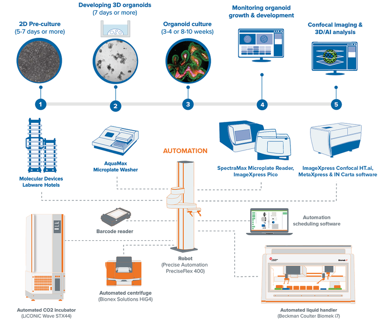
Integrated system for high-throughput intestinal organoid workflow
The automated organoid workcell includes the ImageXpress® Confocal HT.ai High-Content Imaging System, automated CO2 incubator, automated Biomek i7 liquid handler, collaborative robot and rail, and additional, optional instruments including an automated centrifuge, our ImageXpress® Pico Automated Cell Imaging System, and SpectraMax microplate reader. This integrated system allows for automated monitoring, maintenance, and characterization of growth and differentiation of organoids and stem cells, as well as testing the effects of various compounds.
Our lab automation for high-throughput, high-content screening include scientists and engineers who can customize our instruments, as well as automate entire workflows to meet the specific needs of your assay, method, or protocol. From incubators, liquid handlers, and robotics to customized software and hardware—and with over 35 years of experience in the life science industry—you can count on us to deliver quality products and provide worldwide support.
Featured asset
AI-enabled automated compound screening for toxicity effects using healthy human intestinal organoids
This application note highlights a scalable solution that integrates automated organoid culture, compound dosing, and high-content imaging with machine learning–driven phenotypic analysis. By combining deep learning segmentation (SINAP) and phenotypic classification (Phenoglyphs™) found in IN Carta® Image Analysis Software, the system accurately distinguishes between cytotoxic, cytostatic, and viable organoid responses.
Latest resources on organoid research
The complexity of 3D assays remains a hurdle for the wider adoption of organoid models in research and drug screening. New advanced tools for imaging and analysis, as well as assay automation are critical for increased quality of information, throughput, and precision of complex biological models. Learn how high-throughput, high-content imaging and analysis paired with AI-based analytic tools can improve the accuracy of your organoid studies.