
Pulmonary (Lung) Organoids
High-throughput, high-content screening of 3D lung organoids for disease modeling and toxicity assessment
Pulmonary (lung, airway) organoids
Lung organoid cultures are 3D microtissue models recapitulating the morphological and functional characteristics of the airway. They can be formed primary human lung epithelial cells in ECM with growth factors. The cells can self-assemble into the multi-lineage lung epithelium comprising different cell populations.
Lung organoids display characteristic features of the human airway, such as alveolar structure, mucus secretion, ciliary beating, and regeneration. This biological relevance enables the study of repair/regeneration mechanisms in lung injury and phenotypic changes in pulmonary diseases. Lung organoids also can be used for toxicity assessment or drug testing.
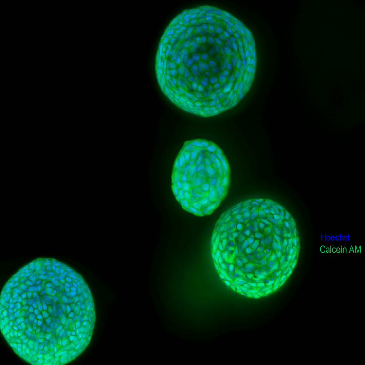
Because lung organoids are hollow, with lumen and cavities inside, they are easily penetrated by light. This makes them compatible with 3D biological assays and suitable for confocal imaging, allowing quantitative characterization of cellular content, live-dead assessment, and cell scoring for specific markers.
Automated cell culture, imaging and analysis of complex biological systems including
Watch our short video with Dr. Oksana Sirenko, senior scientist as she describes an automated integrated system that allows automated monitoring, maintenance, and characterization of growth and differentiation of stem cells and organoids, as well as testing the effects of various compounds.
It demonstrates methods for successful automation and advanced high-content imaging for increased throughput of three complex workflows: iPSC culture, 3D lung organoids, and intestinal organoids.
Automation of 3D lung organoid culture and experimental protocols
Lung organoids can be cultured and monitored via automated workflows. The automated integrated system below includes ImageXpress Confocal HT.ai system and analysis software, automated incubator, Biomek liquid handler, and a robotic device. Machine-learning-based image analysis, can track the growth of diameter and area, density and number of objects. Advanced image analysis allows 3D reconstitution and complex analysis of organoids, including cell morphology, viability, and differentiation markers.
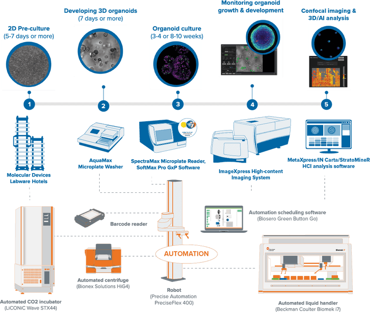
Experimental protocol for lung organoids
- 2D pre-culture - 3D lung organoids were derived from primary human lung epithelial cells and expanded for two weeks in 2D.
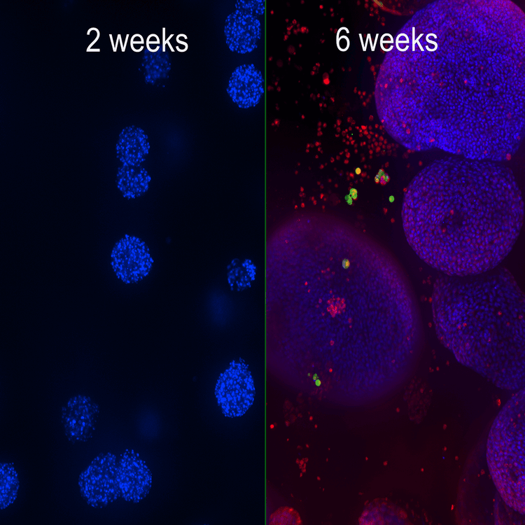
- Developing 3D organoids - Then the cells were seeded in 90% Matrigel domes using reagents. Cells formed 3D organoids and were fed every second day for two weeks using seeding media, then differentiated for another six weeks.
- Organoid culture - Organoids were grown in different plate formats and then treated with compounds or stained with various markers for morphology and viability assessment.
- Monitoring organoid growth & development - Organoid growth and development were monitored in Matrigel using transmitted light imaging or using Hoechst stain that allowed evaluation of organoids size, volume, and complexity.
- Confocal imaging and 3D/AI analysis - Confocal imaging in combination with 3D analysis allowed quantitative characterization of cellular content as well as count and measurement cells with different phenotypes within organoids (cell count, live-dead assessment, cell scoring for specific markers, others). The model can be used for toxicity evaluation of pharmaceutical drugs and other compounds.
Our lab automation for high-throughput, high-content screening include scientists and engineers who can customize our instruments, as well as automate entire workflows to meet the specific needs of your assay, method, or protocol. From incubators, liquid handlers, and robotics to customized software and hardware—and with over 35 years of experience in the life science industry—you can count on us to deliver quality products and provide worldwide support.
Featured asset
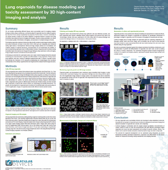
Lung organoids for disease modeling and toxicity assessment by 3D high-content imaging and analysis
In this application, primary human lung epithelial cells were cultured to model human lungs with the formation of 3D lung organoids. The cells within the organoids self-organize into complex structures that retain clusters of multi-lineage epithelial cells.
High-content imaging was used to monitor the growth and differentiation of lung organoids. 3D reconstruction allowed for further complex analysis of the organoid structure. Within the organoids, the cell morphology, viability status, and the expression of various cellular markers can also be measured.
Lung organoid cell image gallery
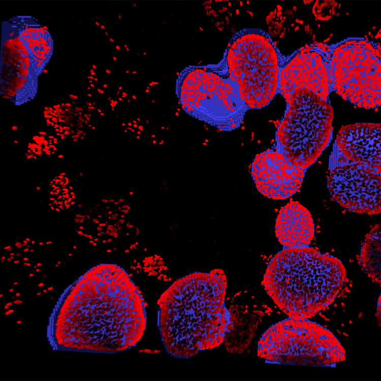
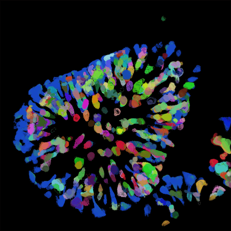
Image analysis of organoid
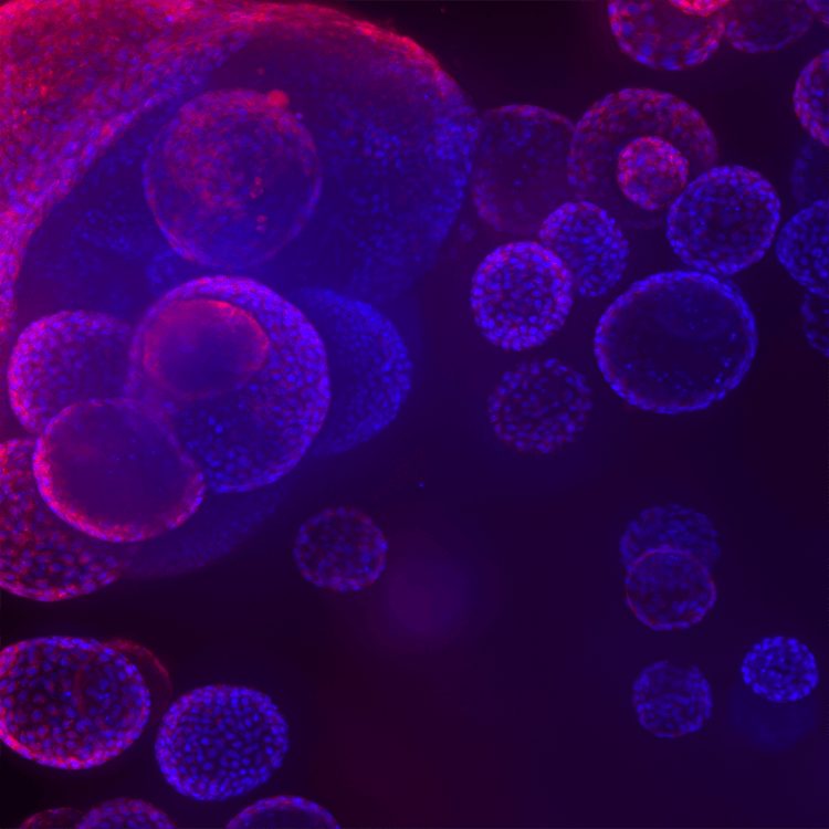
Lung organoids
Lung organoids 2
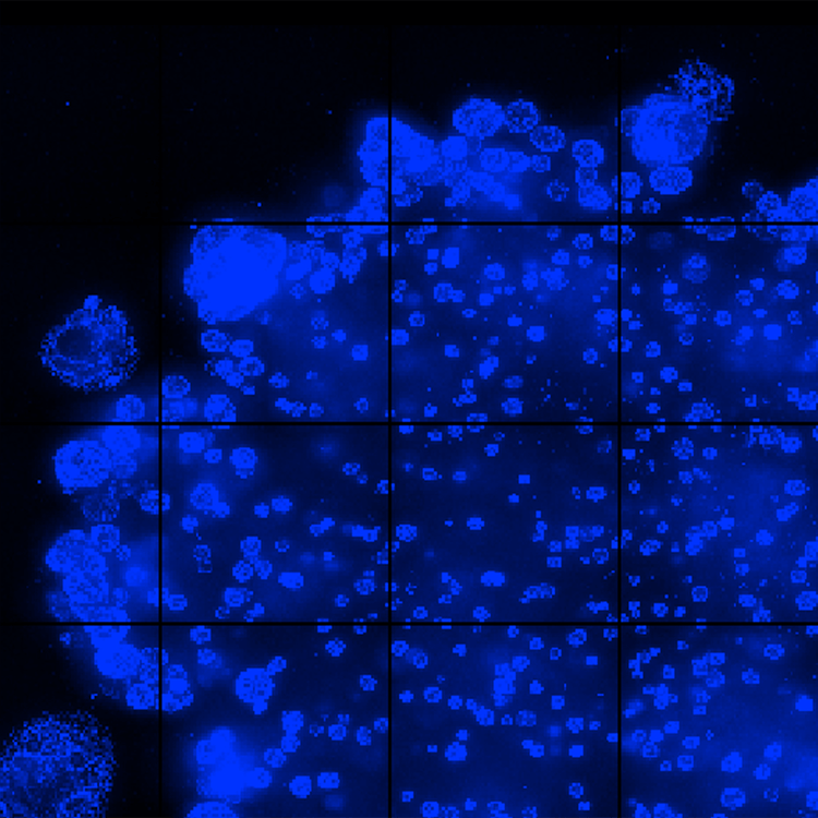

Automated imaging of live organoids in Matrigel was done using confocal option, 4x or 10x magnification
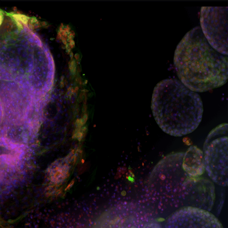

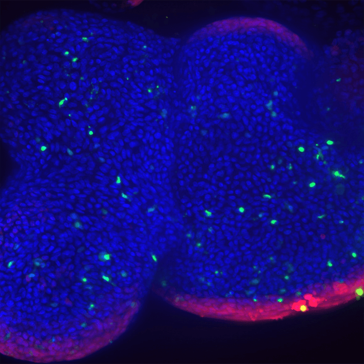
High complexity lung organoid
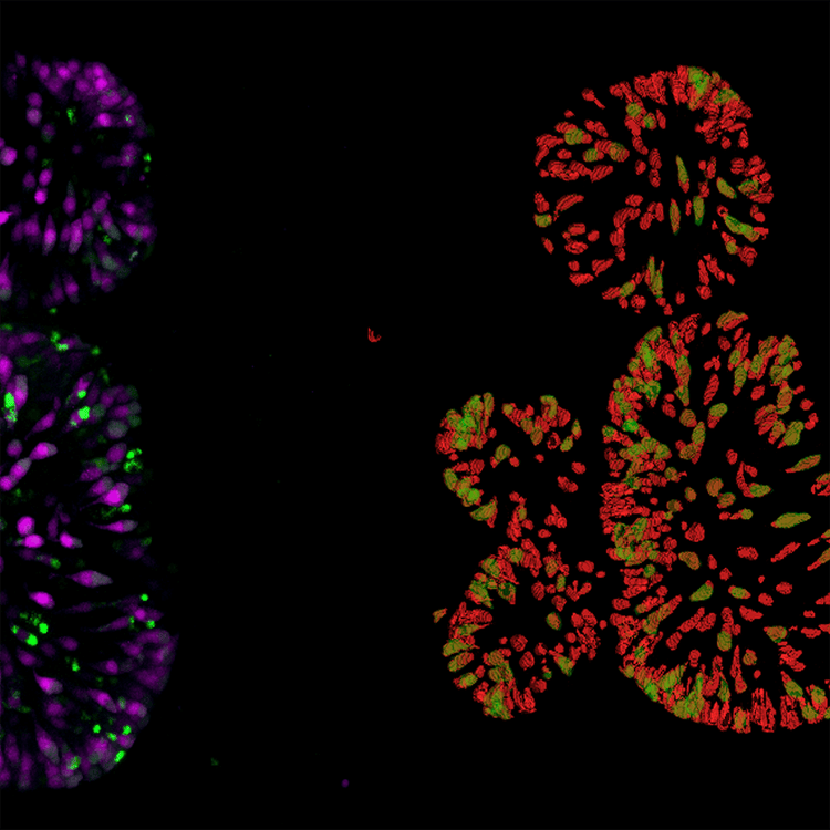
Gut organoid dTomato cells in red Detection of Double positive(mNeon) in green
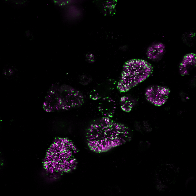
Internally expressed genes dTomato magenta mNeon green 10x
Intestinal organoid 40x,dTomato cell count
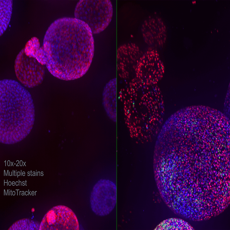
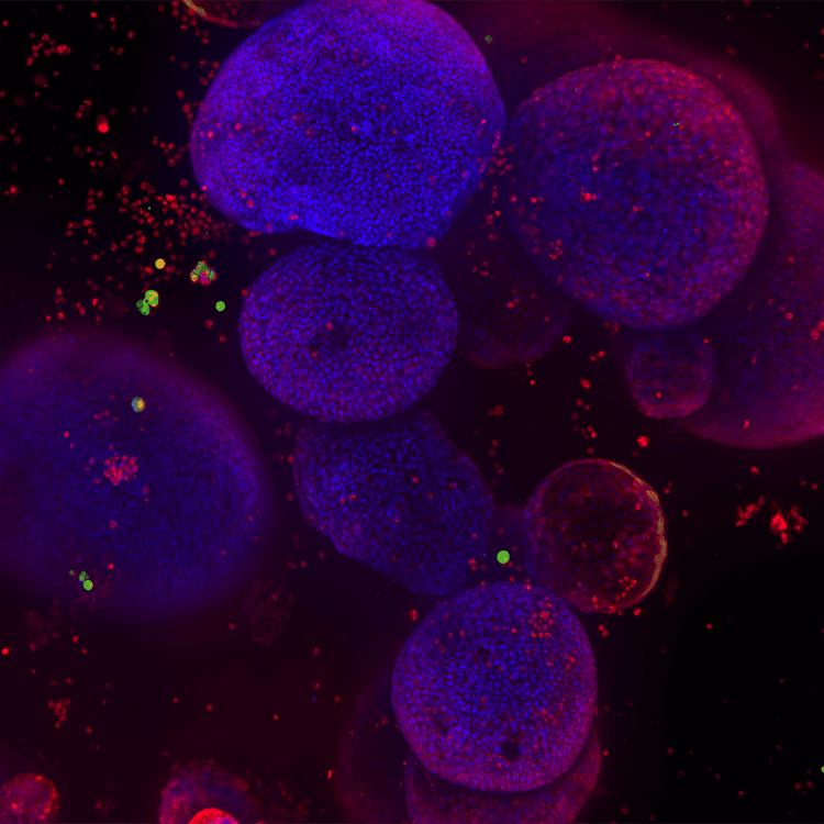
Lung organoid 10x 20x Multiple stains Hoechst MitoTracker
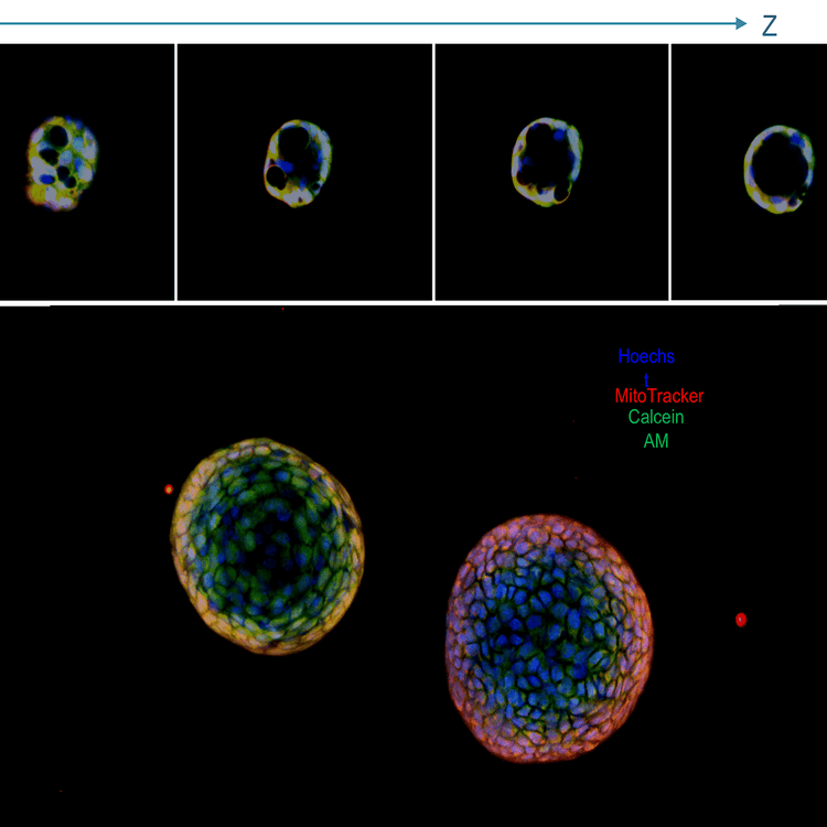
Lung organoids,projection image and a panel with Z stack; live stained with Hoechstblue,nuclei),MitoTracker(red,mitochondria),and Calcein AM(green,viability dye);20x water immersion
Lung organoids 3
Lung organoids 4
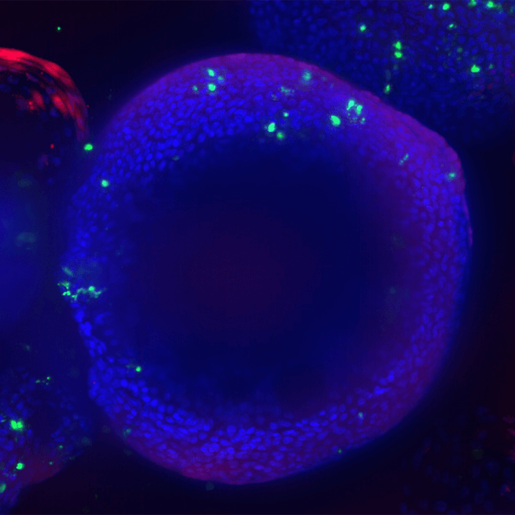
Lung organoids lung airway epithelial cells cultured in matrigel domes for 8 weeks
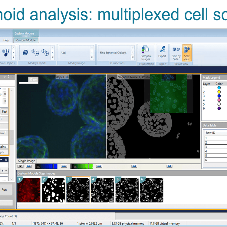
Organoid analysis multiplexed cell scoring
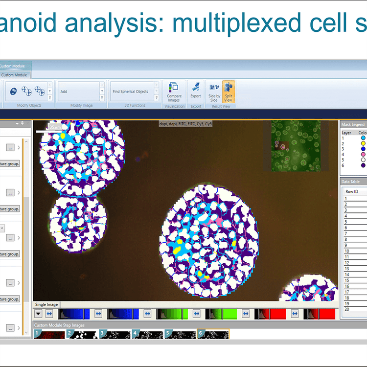
Organoid analysis multiplexed cell scoring 2
Organoid development
Latest resources on organoid research
The complexity of 3D assays remains a hurdle for the wider adoption of organoid models in research and drug screening. New advanced tools for imaging and analysis, as well as assay automation are critical for increased quality of information, throughput, and precision of complex biological models. Learn how high-throughput, high-content imaging and analysis paired with AI-based analytic tools can improve the accuracy of your organoid studies.