Cellular Imaging and Analysis for 3D Cell Culture Applications and Assays
3D cell imaging and analysis workflow
While the development of quantitative assays using 3D cell cultures has become an attractive investigative tool to understand complex biology, challenging 3D cell image acquisition and analysis workflows have hindered wider adoption by the screening community. High-content, high-throughput tools for microscopy offer innovative and automated techniques for evaluating this complex biology. After the 3D cell culture is grown or transferred to an imaging microplate, cells can be treated with compounds and stained with the marker of choice. For acquisition, the ImageXpress® automated imaging systems and our cellular imaging and analysis software provide the capability to screen and analyze these models within a single, integrated interface to dramatically reduce time to discovery.
Steps used for a typical 3D cell culture assay:
3D cell culture imaging and analysis techniques
Our imaging technology continues to evolve for easier implementation of more physiologically-relevant models. Learn more about how our technology can help scale-up 3D cell culture from small-scale assays to high-throughput screening.
Acquire images for 3D assays
In recent years, there have been significant advances in automated microscopy and imaging for delivering more predictive, physiologically-relevant measurements for drug discovery and environmental toxicity applications. Implementation of more complex assays and 3D models requires high resolution to capture publication-quality images and data.
Confocal imaging provides a high signal-to-noise ratio by reducing out-of-focus light for crisper images to yield more cellular detail even within a thick sample. Watch the video to learn about high-throughput techniques on how to acquire images in 3D using the ImageXpress Micro Confocal system.
Analyze images for 3D applications
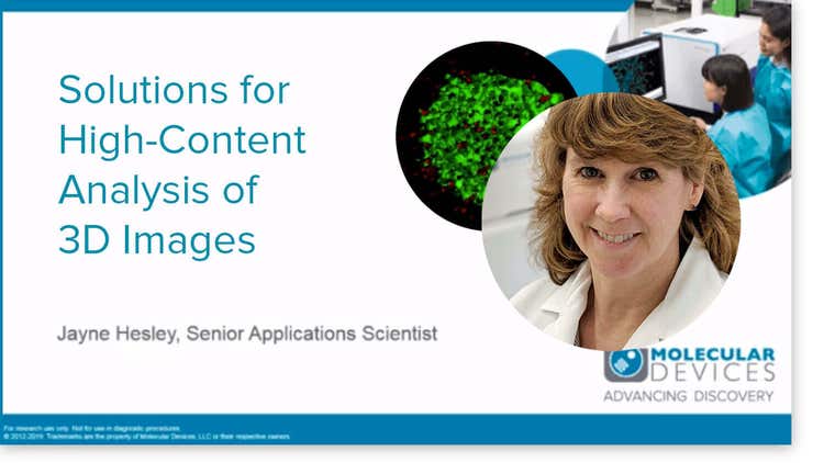
There are different types of image sets that may be generated even though your sample is 3D. A single plane may be sufficient or multiple planes may be acquired. When multiple planes are acquired, it’s useful to be able to analyze them individually or to analyze them after combining them into a single 2D projection. Ultimately, analysis of the entire 3D image may be the goal of the experiment. Watch the video to learn more about techniques for analyzing images from multiple z-planes as a 3D volume using MetaXpress software.
Advantages of 3D Cell Models
Three-dimensional (3D) cell models are more physiologically relevant than two-dimensional cell cultures, and they more closely represent the tissue microenvironments, cell-to-cell interactions, and biological processes that occur in vivo. Now you can generate more predictive data using high-content imaging (HCI) systems like the ImageXpress system. By integrating both high-content image acquisition and analysis software like MetaXpress® software with the 3D Analysis Module, the ImageXpress system can be used for in-depth analysis, visualization, and assessment of 3D structures. This single interface will enable you to meet 3D acquisition and analysis challenges without compromise to throughput or data quality, giving you confidence in your discoveries.
Learn more about 3D cell model application and assays:
-
3D Cancer Cell Research
Cancer involves changes which enable cells to grow and divide without respect to normal limits, to invade and destroy adjacent tissues, and ultimately to metastasize to distant sites in the body. Cancer spheroids mimic tumor behavior far more effectively than standard 2D cell cultures. Such 3D spheroid models are being successfully used in screening environments for identifying potential cancer therapeutics.
Cancer researchers need tools that enable them to more easily study the complex and often poorly understood interactions between cancerous cells and their environment, and to identify points of therapeutic intervention.
3D neural spheroids
To accelerate the development of effective and safe drugs, there is an increasing need for more complex, biologically relevant, and predictive cell-based assays for drug discovery and toxicology screening.
We show that functional and morphological assays using 3D neural spheroids formed with human iPSC-derived cells can be used for evaluation of drug candidates and neurotoxicity assessment.
-
Cardiotoxicity
Cardiotoxicity or cardiac toxicity is a term used to define chemicals that are toxic to the heart, causing muscle damage or heart electrophysiology dysfunction. The assessment of cardiotoxicity is important in the early stages of drug discovery to eliminate potentially toxic compounds from further development. Therefore, there is a growing need for highly predictive in vitro cardiotoxicity assays that use biologically relevant 3D cell-based models and are suitable for high-throughput screening.
Cells in Extracellular Matrices
One common way of culturing cells in three dimensional space is through the use of extracellular matrix-based hydrogels, such as Matrigel. Cells are grown in an extracellular matrix (ECM) to mimic an in vivo environment. Differences between Matrigel and 2D cell cultures can be readily seen by their different cell morphologies, cell polarity, and/or gene expression. Hydrogels can also enable studies on cell migration and 3D structure formation, such as endothelial cell tube formation in angiogenesis studies.
-
Disease modeling using novel flowchip system
Disease Modeling with 3D Cell-Based Assays Using a Novel Flowchip System and High-Content Imaging
There is an increasing interest in using three-dimensional (3D) cell structures for modeling tumors, organs, and tissue to accelerate translational research. We describe here a novel automated organoid assay system (the Pu·MA System) combined with microfluidic-based flowchips that can facilitate 3D cell-based assays.
Organoid Innovation Center
The new Organoid Innovation Center at Molecular Devices combines cutting-edge technologies with novel 3D biology methods to address key challenges of scaling complex 3D biology.
The collaborative space brings customers and researchers into the lab to test automated workflows for organoid culturing and screening, with guidance from in-house scientists.
-
Organoid technology / Organ-on-a-Chip
Organoid technology, like organ-on-a-chip emulates organ physiology though co-culture of cells in a supportive 3D matrix and use of microfluidic channels to perfuse nutrients or compounds over the resulting cellular structures. It is rapidly gaining popularity as a biologically relevant screening model for new drugs and toxicity.
- 3D image analysis and characterization of angiogenesis in organ-on-a-chip model
- Video: Physiologically-Relevant Tissue Models Using a High-Throughput Organ-on-a-Chip Platform
- High-content assay for morphological characterization of 3D neuronal networks in a microfluidic platform
- Webinar: Developing high-throughput organ-on-a-chip tissue models for drug discovery using high-content imaging
- Poster: Water immersion objectives for automated high-content imaging to improve precision and quality of complex biological assays
Organoid vs. Spheroid
The terms spheroid and organoid are often used interchangeably. Both cell models are 3D multi-cellular structures that better mimic the in vivo environment as compared to their 2D cell counterparts. Organoids typically consist of a co-culture of cells which demonstrate a higher order of self-assembly to allow for an even better representation of complex in vivo cell responses and interactions. In contrast, spheroids have a more simplified 3D structure making them amenable to high reproducibility and scaling. Functionally, both model types are used as better in vitro models for capturing more physiologically relevant data.
-
Organoids
Organoids are three-dimensional (3D) multi-cellular microtissues that are designed to closely mimic the complex structure and functionality of human organs. Organoids typically consist of a co-culture of cells which demonstrate a high order of self-assembly to allow for an even better representation of complex in vivo cell responses and interactions, as compared to traditional 2D cell cultures.
Spheroids
Spheroids are multi-cellular 3D structures that mimic in vivo cell responses and interactions. They can be highly reproducible and to be scaled for high-content screening. Compared with adherent cells grown in 2D monolayers, 3D growth conditions are believed to more closely reflect the natural environment of cancer cells. Acquiring measurements from these larger structures involve acquiring images from different depths (z-planes) within the body of the spheroid and analyzing them in 3D, or collapsing the images into a single 2D stack before analysis.
-
Stem Cells
Stem cells are an accessible source of primary-like cells which can be used in longer-term studies to measure targeted responses in specific cell types and 3D tissues. Complex assays and 3D induced pluripotent stem cell (iPSC)-derived cell models better represent tissue biology and cell interactions which make them more relevant for many toxicity and drug screening assays.
Toxicology
Toxicology is the study of adverse effects of natural or man-made chemicals on living organism. It is a growing concern in our world today as we are exposed to more and more chemicals, both in our environment and in the products we use.
-
Whole Organisms
Model systems such as Drosophila, C. elegans, and zebrafish offer relevance to human biology, with the added advantage of genetic and whole-organism phenotypic screening. Zebrafish screening has gained favor as an alternative to mammalian screening due to reduced cost and space requirements, short generation times, and ease of genetic manipulation. Our zebrafish studies use the ImageXpress Micro System and MetaXpress software to set up and run automated imaging screens to monitor inhibition of angiogenesis, quantitate gene knockdown, and measure ototoxicity and neurotoxicity.
3D cell imaging and analysis workflow
The workflow of a 3D cell culture protocol is dependent on the type of cell model being cultured. The 3D cell imaging and analysis workflow depicted here is a generalized example of 3D spheroids in a 96- or 384-well flat or round-bottom microplate. Each step breaks down the process from cell culture to image analysis and highlights the instrumentation and tools needed to conduct the cell culture protocol including an automated high-content imaging system and cellular imaging and analysis software.

CULTURE SPHEROIDS
3D cell can be cultured directly in a ULA, round bottom plate to develop the typical spherical shape of a spheroid. Cells can also be grown in alternate labware like a petri dish, flask, or other microplate before transferring to an imaging microplate.

96- or 384- Well Plates
Most 3D cell cultures for high-content, high-throughput screening are run using 96- or 384-well microplates. ULA, U-shaped/round bottom microplates enable the spherical shape to form spheroids or spheroids can also be cultured in a flat-bottom microplate using hydrogels to simulate extracellular matrices (ECM). Optimized spheroid cell culture protocols for the ImageXpress® Micro Confocal High-Content Imaging System can use ULA, U-bottom 96- and 384-well plates with a one-step staining procedure that reduces assay time and minimizes variability.
Shopping for reagents?
Check out our
EarlyTox Cell Viability Assay Kits


TREAT WITH COMPOUNDS
After spheroid formation, compounds are added into the wells, and then incubated for one to several days, depending on the mechanism being studied. Generally, shorter durations are used to study apoptosis and longer durations for multi-parameter cytotoxicity studies. For drug treatments requiring a longer duration, compounds are refreshed periodically during incubation. The concentration of compounds and the incubation period is dependent on the desired protocol.

STAIN FOR MARKERS
After compound treatment is completed, stains are added directly to the media. Stains that require no washing can be used to avoid disturbing spheroids, but spheroids can be carefully washed even using automation, if necessary.

Assay Kits
Easy-to-use, robust assay kits for life science research, drug discovery and development, and bioassays. Our assay kits are optimized for use on our instruments. Screen more compounds earlier in drug discovery and enable characterization of a full concentration-response profile of test compounds for cell viability, cell proliferation, axiogensis, and more.

High-content Imaging System
The ImageXpress Micro Confocal system is a unique confocal imaging solution capable of imaging more than a million wells a week. MetaXpress software enables acquisition of 3D z-stacks. Z-plane images can be saved individually or collapsed into a single 2D projection image using a mathematical algorithm.

ACQUIRE 3D CELL IMAGE
Images within the body of the spheroid can be captured individually or as a z-stack (multiple images taken at differing depths) using specialized imaging equipment.

ANALYZE 3D CELL IMAGE
Use cellular imaging analysis software to run quantitative analysis of the cell images to assess and monitor how cells express against different markers and to quantify biological readouts.

High-content Imaging Software
MetaXpress High-Content Image Acquisition and Analysis Software is a multi-level analysis tool for a wide range of 2D and 3D applications optimized for the ImageXpress Micro Confocal system. The integrated acquisition and analysis application modules for 3D cell models simplify high-throughput quantification of 3D structures with volume, intensity, and distance measurements.
Resources for 3D Cells
Blog
3D organoids and automation of complex cell assays [Podcast]
As we enter the era of sophisticated drug discovery with gene therapy and personalized medicine, we need to be prepared to study complex diseases, assess the therapeutic effect of…
Application Note
Monitoring of T-cell invasions assay using a 3D spheroid model
T-cell therapies are designed to help our immune system eliminate cancer cells. Those include CAR T-cells (Chimeric Antigen Receptor engineered T-cells), tumor infiltrating lymphocytes (TIL…
Publications
Gene editing in organoids: accounting for complexity in drug discovery
More researchers are using gene editing to build disease models that better represent human tissues' complex biology, signaling a shift away from 2D cell culture or animal models to organoid…
Publications
Bioprinting Automation for Drug Discovery to Be Developed by Molecular Devices and Advanced Solutions
3DPrint.com, the leading source for actionable intelligence related to 3D printing technology and the larger AM industry, covers our collaboration with Advanced Solutions to develop 3D biolo…
Publications
Improving Drug Development: Molecular Devices and Cellesce Aim to Advance the Use of Organoids
Following our acquisition of Cellesce Ltd, Tanya Samazan from Instrument Business Outlook dove deep into conversation with Molecular Devices President Susan Murphy and Cellesce CEO Vicky Mar…
News
Molecular Devices and Advanced Solutions Life Sciences collaborate to develop 3D Biology Automation Technologies for Drug Discovery
Turnkey platform integrates flexible robotic automation with high-content imaging of complex 3D cellular models, enabling high-volume organoid screening SAN JOSE, Calif., Jan. 4, 2023 – Mole…
Application Note
Imaging characterization of a 3D bioprinted ovarian cancer model
3D bioprinting is defined as the additive deposition of cells and biocompatible materials to build biologically functional 3D structure or artificial tissue models
Brochure
BioAssemblyBot 400 (BAB400): more than a bioprinter
The BioAssemblyBot® 400 is an intelligent robot used by life scientists to build 3D model systems with increased throughput and precision, alleviating major concerns mentioned above.
News
Molecular Devices adds proprietary patient-derived organoid technology with acquisition of Cellesce
First-of-its-kind technology from Cellesce creates consistent patient-derived organoids for large scale drug screening Acquisition strengthens Molecular Devices’ position as a 3D biology sol…
Publications
Overcoming ‘Analysis Paralysis’ in Cell Painting With Artificial Intelligence
Gone are the days of only measuring single parameters in cell-based experiments. Instead, researchers should widen their focus, namely with the help of innovations like the Cell Painting ass…
Application Note
Automated dispensing, monitoring, and assay development of hydrogel-based cellular models
3D cellular models that more accurately represent various microenvironments are incredibly important for accurate drug screening and disease modeling.
Scientific Poster
Automation for organoid assays – An integrated system with high-content imaging
Three-dimensional (3D) cell models that represent various tissues are being successfully used in drug discovery and disease modeling to study complex biological effects and tissue architectu…
Publications
How cardioids can usher in the next generation of drug discovery
We recently partnered with HeartBeat.bio to automate and scale the production of cardioids — 3D cell models of the heart that can more accurately recapitulate human biology. Labiotech.eu spo…
Publications
What an innovation centre means for 3D biology
In April 2021, we launched our Organoid Innovation Centre (OIC), an initiative designed to help scientists leverage the potential of 3D biology throughout the drug discovery process. On our…
eBook
Building complexity in organoid models
Building complexity in organoid models | Molecular Devices
Publications
Q&A: What the transition away from animal testing could mean for drug discovery
In September, the U.S. Senate unanimously passed the FDA Modernization Act 2.0, which would lift an 84-year-old federal mandate for animal testing for toxicity studies. While the bill doesn’…
Blog
Advanced technology for automated 3D biology workflows #SLASEurope2022
SLAS Europe 2022, hosted numerous sessions packed with the latest research on emerging topics as well as sessions and panel discussions focused on how to build, and succeed, in a…
News
Molecular Devices expands global R&D hub in Austria
Larger site will be future home of the Organoid Innovation Center – Salzburg, a collaborative space for advancing automated cell line development, organoid development, and screening solutio…
Application Note
Automated Monitoring of Development and Activity Analysis of iPSC-derived 3D Cerebral Organoids
The human brain is highly complex which makes it challenging to study both in vitro and in-model organisms. Cultured neurons do not sufficiently recapitulate the three-dimensional (3D)…
News
Molecular Devices and HeartBeat.bio announce collaboration to automate and scale cardiac organoids for high-throughput screening in drug discovery
SAN JOSE, Calif., and VIENNA, Sept. 12, 2022 – Molecular Devices, LLC., a leading provider of high-performance life science solutions, and HeartBeat.bio AG, a biotech company developing card…
Application Note
AI enabled phenotypic analysis of label-free intestinal organoids
The intestinal crypt system is an important part of the intestinal organoid where mature organoids have more complex and numerous crypt structures. Yet, due to their high turnover rate and…
Blog
Stem Cell Science and Regenerative Medicine – Technology and Methods Presented at ISSCR 2022
It was another great year at ISSCR 2022! Leaders from across the globe came together to discuss new technologies, share insights, and explore the newest breakthroughs in stem cell…
eBook
Capture True 3D Cell Culture Insights
Development of more complex, biologically relevant, and predictive cell-based assays for compound screening is a primary challenge in drug discovery. The integration of three-dimensional (3D…
Application Note
Neurite outgrowth analysis in 3D reconstructed human brain micro-tissues
Organotypic 3D cultures resemble native 3D tissue architecture and are believed more representative of real tissues than their 2D (monolayer) counterparts, thus providing higher in vivo…
Blog
How automated organoid cell cultures are developed, imaged, and analyzed
3D cell models are becoming increasingly popular for studying complex biological effects, tissue functionality, and diseases. Their ability to self-organize and mimic…
Application Note
Automation and high content imaging of 3D triple-negative breast cancer patient-derived tumoroids assay for compound screening
Triple negative breast cancer is a clinically aggressive tumor subtype, with high rates of metastasis, recurrence, and drug resistance.
Application Note
3D Immune Cell Migration
Cell migration is an essential process for many biological phenomena such as early embryonic development and immune cell response. During inflammation, the recruitment of T cells through…
Publications
Benefits of Real-Time Live-Cell Imaging
Three-dimensional (3D) cell-based assays replace a host of tissue, cell, and animal test models but have a reputation for lacking time resolution. For example, traditional, endpoint cell-bas…
Scientific Poster
Automation of the organ-on a chip assay: automated culture, imaging and analysis of angiogenesis
There is a critical need for biological model systems that better resemble human biology. Three-dimensional (3D) cell models and organ-on-a-chip (OoC) structures representing various tissues…
Blog
Life Science and Technology Predictions for 2022
For three decades, Molecular Devices has made scientific breakthroughs possible for academic, pharmaceutical, government, and biotech customers with its innovative life science…
Blog
Engineering Next-gen Organoids with Automated Lab Workflows at #SLAS2022
SLAS2022, the Society for Lab Automation and Screening conference offered another exciting year for learning about innovative laboratory technologies. Whether you attended in-person…
Blog
Enabling 3D High-Content Imaging and Analysis on the Organ-on-a-Chip Platform
Imagine having the ability to mimic the human biological environment for disease modeling and drug screening and doing so in a micro-scale system. With the development of organ-on-a-…
Scientific Poster
Automation of 3D organoid culture workflow with deep-learning based image analysis
3D cell culture as a model system is increasingly popular because it recapitulates the in vivo microenvironment better than 2D cell cultures. Organoids have the capacity for stable different…
Blog
Stem cell science insights and breakthroughs presented at #ISSCR2021
If you didn't get a chance to visit us at our poster sessions during ISSCR 2021, don’t fret. We've gathered all our sessions right here for you. The ISSCR Annual Meeting brought…
Scientific Poster
Organoids for disease modeling and in vitro drug screening
3D cell models representing various tissues were successfully used for studying complex biological effects, tissue architecture, and functionality. However, the complexity of 3D models remai…
Scientific Poster
Monitoring organoid development and characterization of calcium oscillation activities in iPSC-derived 3D cerebral organoids
Cerebral organoids are a rapidly developing technology that has great potential for understanding brain development and neuronal diseases. They can also be used for testing effects of compou…
Scientific Poster
High-throughput assessment of compound-induced pro-arrhythmic effects in human IPSC-derived cardiomyocytes
Development of biologically relevant and predictive cell-based assays for compound screening and toxicity assessment is a major challenge in drug discovery. The focus of this study was to es…
Scientific Poster
Deep learning-based image analysis for label-free live monitoring of iPSC 3D organoid cultures
Complex 3D biological models such as organoids and patient derived spheroids are gaining popularity in many biomedical research areas because they more closely recapitulate the in vivo tissu…
Publications
Project Profile: Organoid Innovation Center
Lab Manager speaks to Dan O’Connor, vice president, drug discovery, Molecular Devices, about the company’s Organoid Innovation Center in San Jose, CA. The center is 180 sq. ft., within a lar…
Publications
Organs-on-Chips: Expand the Boundaries of In Vitro Testing
Humans began culturing animal cells and tissues as early as the late 19th century, when Wilhelm Roux first showed that chick embryos could be cultured in saline solution for a few days. Even…
Blog
Tips for running a successful live cell imaging experiment
There have been significant advancements in microscopy and camera technology, as well as advancements in technologies for labeling molecules of interest over the past decade. These…
Blog
Overcome the challenges of high-throughput 3D imaging
Thanks to recent advances in imaging technologies, we are now able to observe and analyze complex cellular networks in three dimensions. Through 3D imaging, we can acquire and…
Publications
Shaping the Future of Organoid Research
Molecular Devices, a provider of innovative life science technology, recently unveiled a brand new, first-of-its-kind Organoid Innovation Center. Situated at the company’s global headquarter…
Publications
Disease Modeling with 3D Cell-Based Assays Using a Novel Flowchip System and High-Content Imaging
There is an increasing interest in using three-dimensional (3D) cell structures for modeling tumors, organs, and tissue to accelerate translational research. We describe here a novel automat…
Application Note
Lung organoids as an assay model for in vitro assessment of toxicity effects by 3D high-content imaging and analysis
Organoid models have increasingly gained popularity in biologic research and screening to recapitulate complexity of real tissues. To model the in vivo human lungs, we have cultured primary…
Blog
Overcome the challenges of high-content cell analysis through AI/machine learning
Artificial intelligence (AI) is finding its way into many aspects of modern life, from autonomous vehicles to voice-powered personal assistants, and even the creation of art. But it…
Publications
Using 3D cancer cell models to push forward personalized medicine
Using 3D cancer cell models to push forward personalized medicine: an interview with Angeline Lim, PhD, of Molecular Devices Editor-in-Chief, Francesca Lake, speaks to Angeline Lim of Mole…
Publications
Disease Modeling with 3D Cell-Based Assays Using a Novel Flowchip System and High-Content Imaging
There is an increasing interest in using three-dimensional (3D) cell structures for modeling tumors, organs, and tissue to accelerate translational research. We describe here a novel automat…
Blog
How Cell Painting is making its mark on drug discovery
Have you ever heard the old adage, “A picture is worth a thousand words?” When it comes to Cell Painting, this saying is especially true. Cell Painting is a high-content,…
Publications
Danaher’s SCIEX and Molecular Devices Businesses Debut New Products
It has been a busy beginning of the year for Danaher’s Life Sciences segment. In January, the business reported 2020 revenues of $10.6 billion, representing 47% of the total revenues of scie…
Blog
Get to know our Field Applications Scientist: Dwayne Carter
Dwayne Carter gives us a taste of 3D bioprinting, clone screening, and Caribbean cuisine Dwayne Carter is a cell biologist and educator who joined Molecular Devices in November 2020…
Blog
7 tips for optimizing your 3D cell imaging and analysis workflow
As the field of cell biology moves toward increasing complexity of assays, 3D cell systems have proven to provide more physiologically relevant information as compared to…
Blog
Get to know our Field Applications Scientist: Kayla Hill
Kayla Hill discusses the latest trends in cellular imaging We recently hosted a webinar with our Field Applications Scientist, Kayla Hill, PhD, who explored high-content analysis…
Scientific Poster
3D imaging and analysis of angiogenesis in the organ-on-a-chip platform
Angiogenesis is the physiological process of formation and remodeling of new blood vessels and capillaries from pre-existing blood vessels.1 This can be achieved through endothelial sproutin…
Application Note
High-throughput screening of 3D cell cultures with multiple high density scaffold-free spheroids for cancer toxicity studies
3D spheroid models for cancer research are gaining popularity because they better mimic the in vivo tissue architecture, gene expression and metabolic profile of tumors compared to…
Scientific Poster
AI-based analysis of complex biological phenotypes
Cell-based phenotypic assays have become an increasingly attractive alternative to traditional in vitro and in vivo testing in pharmaceutical drug development and toxicological safety assess…
Publications
Improving acquisition and analysis of 3D cell model assays with water immersion objectives
Sebastian Peck is a senior product manager for cellular imaging at Molecular Devices, one of the world's leading providers of high-performance bioanalytical measurement systems, software, an…
Publications
The workflow game-changer: combined technologies for efficiency
Microfluidic flowchip technology combined with three-dimensional (3D) imaging is a powerful duo poised to change the future of drug discovery and development. The integration of combined tec…
Publications
Neuroscience: bridging the gap between cell-based and human research
Neurological disorders impact up to a billion people around the world and appear to be on the rise [1]. In fact, those affecting the central and peripheral nervous systems – including stroke…
Application Note
3D image analysis and characterization of angiogenesis in organ-on-a-chip model
Angiogenesis is the physiological process of formation and remodeling of new blood vessels and capillaries from pre-existing blood vessels. This can be achieved through endothelial…
Application Note
Phenotypic characterization of neuroactive compound effects
The FLIPR Penta system is powered by a high-speed EMCCD camera and the new ScreenWorks Peak Pro 2 software. The system allows measurement and analysis of complex patterns of calcium…
Scientific Poster
Multiplexed automated assays for neurotoxicity evaluation using induced pluripotent stem cell-derived neural 3D cell models
Cell-based phenotypic assays have become an increasingly attractive alternative to traditional in vitro and in vivo testing in pharmaceutical drug development and toxicological safety assess…
Publications
3D Microscopy Keeps Getting Faster, Smarter, Leaner
With each passing day, continuous advancements are being made in the studies that cutting-edge instruments can perform and in the complexity of the biological samples that are being examined…
Publications
From 3D high throughput to simple automated imaging: Cellular imaging within reach of every laboratory
OUR ABILITY to image cells has come a long way since the pioneering days of Galileo Galilei and Antonie van Leeuwenhoek. The multiple imaging techniques available today range from simple lig…
Publications
Making the move from 2D to 3D
Jayne Hesley is a Senior Applications Scientist for Cellular Imaging at Molecular Devices, LLC. She has over 10 years’ experience developing cell-based applications using ImageXpress Micro h…
Publications
Revolutionizing early drug discovery for immuno-oncology and neurodegenerative disease modeling
The development of 3D cellular models of disease has enabled researchers to better recapitulate in vivo cell environments. As 3D models become more prevalent in the drug discovery industry,…
Scientific Poster
Water immersion objectives for automated high-content imaging to improve precision and quality of complex biological assays
The purpose of these studies was to determine if water immersion objectives, used to improve image quality in complex biological assays, could be used in a high-throughput environment.
Publications
2020 Society for Lab Automation and Screening International Conference & Exhibition: Laboratories Embrace Automation
The Society for Laboratory Automation and Screening (SLAS) held their annual conference January 25–29 in San Diego, California. The show hosts presentations, short courses and an exhibition…
Application Note
Functional evaluation of neurotoxic and neuroactive compound effects in iPSC-derived 3D neural co-cultures
There is increasing interest in using more complex, biologically-relevant, and predictive cell-based platforms for assay development and compound screening.
Presentations
Functional and mechanistic neurotoxicity profiling using human iPSC-derived neural 3D cultures
Expertise in human iPSC-derived neural and cardiac platforms; Focus on adapting platforms to HTS and producing assay-ready solutions to accelerate drug discovery; Neural platform: mocroBra…
Publications
Functional and Mechanistic Neurotoxicity Profiling Using Human iPSC-Derived Neural 3D Cultures
Neurological disorders affect millions of people worldwide and appear to be on the rise. Whereas the reason for this increase remains unknown, environmental factors are a suspected contribut…
Application Note
Assay apoptosis in magnetically bioprinted HepG2 spheroids
Learn how HepG2 cells can be bioprinted using a magnetic 3D cell culture system and imaged on the SpectraMax i3x plate reader with MiniMax 300 Imager.
eBook
Acquire and analyze 3D images like a pro
There has been significant progress in the development of 3D models and techniques during the last few years. Methods include biodegradable scaffolds, organ-on-a chip structures, or self-ass…
Application Note
Acquire and analyze images of FUCCI spheroids on the SpectraMax MiniMax cytometer
Spheroids are small three-dimensional (3D) cellular microenvironments grown using a variety of specialized culture methods such as low-adhesion microplates. This 3D cell culture confers a…
Scientific Poster
Novel Imaging and Analysis of Zebrafish for High Throughput Screening
Explore noval imaging & analysis of Zebrafish for high throughput screening, where Zebrafish Is an attractive model system for developing disease related assays for drug discovery.
Application Note
Measuring mitochondrial shape and distribution using the ImageXpress Micro High-Content Imaging System
Using high-content imaging assays allow cells cultured and treated in microplates to be imaged alive without wash steps for real-time studies of cells in their most natural state, although…
Application Note
Assessment of drug effects on cardiomyocyte physiology using human iPSC-derived cardiac spheroids
Cell models are becoming more complex in order to better mimic in vivo microenvironments and provide greater predictivity for compound efficacy and toxicity. There is an increasing interest…
Application Note
High-content assay for morphological characterization of 3D neuronal networks in a microfluidic platform
Establishment of physiologically-relevant in vitro models is crucial to further understanding of the mechanisms of neurological diseases as well as targeted drug development. While iPSC-…
Application Note
Automating the generation and analysis of 3D cell cultures in Matrigel®
Researchers are turning to three-dimensional (3D) cell culture as a way to increase the biological relevance of their disease models and predictive value of drug studies. These 3D models…
Application Note
Multi-parameter imaging assay for measuring toxicity in a tumor model
There is an increasing interest in using three-dimensional (3D) spheroids for modeling cancer and tissue biology to accelerate translational research. The goal of this study was to develop…
Application Note
3D analysis and morphometric characterization of compound effects on cancer spheroid cultures
Cellular transformation/tumorigenicity assays using cultures of cells in semi-solid media (soft agar or Matrigel) has been well established for cancer research1,2,5. The assay requires…
Application Note
High-content 3D toxicity assay using iPSC-derived hepatocyte spheroids
There is increasing interest in exploring the use of three-dimensional (3D) spheroids for modeling developmental and tissue biology with the goal of accelerating translational research in…
Application Note
High-Throughput Confocal Imaging of Spheroids for Screening Cancer Therapeutics
In recent years, there has been significant progress in development of in vitro aggregates of tumor cells for use as models for in vivo tissue environments. When seeded into a well of a…
Application Note
3D Imaging of Cancer Cell Spheroids
Many cancer cell lines will form spheroids if cultured on a favorable three dimensional (3D) matrix. These spheroids are believed to represent tumor physiology more closely than cells…
Application Note
Analyze cardiomyocyte spheroid contractions with SoftMax Pro Software
Cell-based models for compound screening have become more complex in order to represent complexity of biological systems. Live-cell imaging and threedimensional models provide valuable…
Application Note
High-throughput imaging assays using zebrafish, a model organism for human disease
Recently, zebrafish-based screening has gained favor as an alternative to mammalian screening due to cost, throughput and reduced ethical concerns. Zebrafish are a useful model for drug…
Application Note
High-content screening of neuronal toxicity using iPSC-derived human neurons
The nervous system is sensitive to the toxic effects of many chemical compounds, environmental agents and certain naturally occurring substances. Neurotoxicity can cause temporary or…
Application Note
Multiplexed high-content hepatotoxicity assays using iPSC-derived hepatocytes
Drug-induced hepatotoxicity is an important cause for liver injury and acute liver failure. Thus highly predictive assays for safety and efficacy testing are crucial for improving drug…
Scientific Poster
High-Content Assay for Morphological Characterization of 3D Neuronal Networks in a Microfluidic Platform
The absence of physiologically relevant in vitro models of the nervous system is an important limitation in using 3D cultures for assay development applicable for neurodegenerative diseases…
Scientific Poster
In Vitro Assessment of Drug Effects on Human iPSC-Derived Cardiac Spheroid Cultures
for modeling developmental and tissue biology with the goal of accelerating translational research. Such 3D models can provide different perspectives from traditional 2D cultures on the resp…
Publications
Phenotypic Assays for Characterizing Compound Effects on Induced Pluripotent Stem Cell-Derived Cardiac Spheroids
Development of more complex, biologically relevant, and predictive cell-based assays for compound screening is a major challenge in drug discovery. The focus of this study was to establish h…
Publications
Phenotypic Characterization of Toxic Compound Effects on Liver Spheroids Derived from iPSC Using Confocal Imaging and Three-Dimensional Image Analysis
Cell models are becoming more complex to better mimic the in vivo environment and provide greater predictivity for compound efficacy and toxicity. There is an increasing interest in explorin…
Publications
High-Content Assays for Characterizing the Viability and Morphology of 3D Cancer Spheroid Cultures
There is an increasing interest in using three-dimensional (3D) spheroids for modeling cancer and tissue biology to accelerate translation research. Development of higher throughput assays t…
Videos & Webinars

Organoid Innovation Center Walkthrough

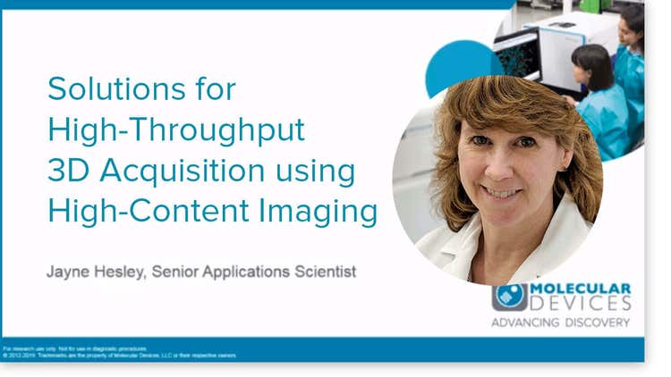
Solutions for High-Throughput 3D Acquisition using High-Content Imaging

Solutions for High-Content Analysis of 3D Images

Zebrafish Actin mask w FITC vessel mask

Angiogenesis Research: High-Content Imaging Systems Help Unlock the Full Potential of 3D Tissue Models

Physiologically-Relevant Tissue Models Using a High-Throughput Organ-on-a-Chip Platform