

ImageXpress Confocal HT.ai High-Content Imaging System [Discontinued]
A scalable, high-throughput, high-content screening solution with 7-channel high-intensity laser light source and machine learning capabilities.
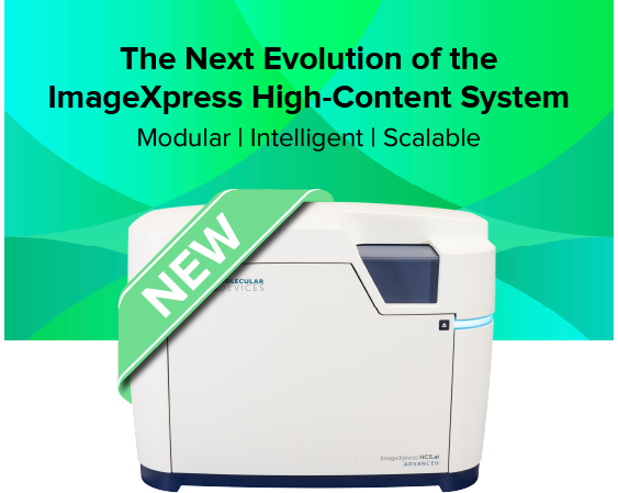
Introducing ImageXpress HCS.ai High-Content Screening System — Modular, intelligent, and built for the future of high-content imaging
Building on the trusted legacy of the ImageXpress Confocal HT.ai system, Molecular Devices introduces the next evolution in high-content imaging — the ImageXpress HCS.ai High-Content Screening System. This fifth-generation modular platform is designed to scale effortlessly with your research, empowering you to move seamlessly from widefield imaging to advanced laser-based confocal modalities—all within one adaptable system.
Engineered for discovery without limits, ImageXpress HCS.ai system combines the renowned optical excellence you trust with revolutionary new capabilities: accelerated acquisition speeds, the intuitive, redesigned MetaXpress® Acquire software, and next-generation, AI-driven IN Carta® analysis to extract deeper insights from complex 2D and 3D models.
Whether you're progressing from widefield microscopy to confocal or expanding into advanced, AI-powered 3D analysis, the ImageXpress HCS.ai system delivers the speed, flexibility, and intelligence to keep your research at the cutting edge—built upon the innovation you already trust.
ImageXpress Confocal HT.ai: Capture large 3D organoid and spheroid images with up to double the speed
The ImageXpress® Confocal HT.ai High-Content Imaging System utilizes a seven-channel laser light source with eight imaging channels to enable highly multiplexed assays while maintaining high throughput by using shortened exposure times. Water immersion objectives improve image resolution and minimize aberrations so scientists can see deeper into thick samples.
The powerful combination of MetaXpress® software and IN Carta® software simplifies workflows for advanced phenotypic classification and 3D image analysis with machine learning capabilities and an intuitive user interface.

Enable greater assay flexibility
Eight imaging channels with laser excitation enable more assay flexibility, higher image brightness, and flexibility to use targeted imaging such as QuickID. Automated Water Immersion objectives offer greater numerical aperture and matched refractive index between the sample and the immersion media for enhanced resolution and decreased aberrations.

Increase throughput with higher quality images
Higher excitation power provides increased signal, shorter exposure times, and faster acquisitions of 3D samples. Micro-lens enhanced spinning disk confocal provides a flat field of view for more accurate and reproducible image analysis. Shorter exposure times generates up to a two-fold boost in scan speed. FRET experiments using lasers for CFP and YFP expand research.

Accelerate analysis speeds
IN Carta Image Analysis Software performs complex segmentation and classification. Phenoglyphs provides a robust trainable classification, and SINAP provides trainable segmentation for any image type. Accelerate analysis speeds by 40X with multi-threaded, parallel processing with MetaXpress® PowerCore Software. Reduces time from hours to minutes, eliminating 3D analysis as a bottleneck.
Introduction to the Organoid Innovation Center and ImageXpress Confocal HT.ai system
Features

High-intensity laser light source
Capture images faster with shorter exposure times. Multiplex your experiments with 7 lasers and 8 imaging channels.

Accurate 3D measurements
Custom Module Editor 3D analysis is optimized for confocal imaging, enabling 3D measurements of volume and distance.

Wide field of view
Wide field of view enables whole-well confocal imaging and eliminates missed targets. Next-generation dual microlens enhanced spinning disk technology provides a large, flat field of view for more accurate and reproducible analysis.

Exclusive AgileOptix™ spinning disk technology
Provides increased sensitivity with specially designed optics, high-powered laser illumination, and sCMOS sensor. Swappable disk geometries provide flexibility between speed and resolution.

IN Carta Image Analysis Software
Leverages machine learning to improve the accuracy and robustness of high-content image analysis, delivering data insights that other technologies miss. Reduces the complexity of image analysis with intuitive guided workflows in a modern user interface.

Multiple imaging modes
The system offers phase contrast and brightfield label-free imaging, widefield and confocal fluorescent imaging with water immersion optics as a standard option.

Automated water immersion objective technology
Offers greater image resolution and sensitivity with up to 4x increase in signal leading to lower exposure times.

Wide dynamic range
Quantifies low and high intensity signals in a single image with >3 log dynamic range intensity detection.
Images taken with same exposure times show different average intensities
IN Carta image analysis software
Powerful analytics combined with an intuitive user interface simplify workflows for image analysis and phenotypic profiling. Advanced features provide the functionality you need to analyze data in 2D, 3D, and 4D - at scale - and deliver real-time insights without the need for complex pre- or post- processing operations. Improve specificity of your image analysis workflows by utilizing the SINAP deep-learning module and see for yourself that Segmentation Is Not A Problem. Put machine learning to work for you and perform complex phenotypic analysis within a user-friendly Phenoglyphs module.

Cellular Image Gallery

HT.ai Organoid with Black Hole

HT.ai Organoid overlay z-steps

HT.ai Cell Painting

HT.ai Organoid-3

HT ai organoid encoded
†Data and images were acquired during development using customer samples. Results may vary. Highlighted features’ price, time to deliver, and specifications will vary based on mutually agreed technical requirements. Solution requirements may cause adjustment to standard performance.
Latest Resources
Featured Applications
Customer Breakthrough
Expand your research using flexible, high-performance imaging solutions
Molecular Devices provides flexible options for the ImageXpress Confocal HT.ai system to meet your research needs and to easily capture images from different sample formats, including hanging drops and in round or flat bottom plates, for monitoring cell health kinetics under environmental control, and more. With over 30+ years of imaging expertise, we can help you select the right options to ensure the best images for your assay.
Standard hardware options

Water immersion objectives
20X, 40X, and 60X water immersion objectives improve the geometric accuracy during acquisition and reduce light refraction for brighter intensity at lower exposure times.

Transmitted light tower
Our transmitted light tower enables acquisition of high contrast images for unstained cells which can be easily viewed or separated from background.

Environmental Control
Environmental control maintains temperature and humidity levels while minimizing evaporation for multi-day, live cell, time-lapse imaging.
Customization options
Molecular Devices can successfully tailor the ImageXpress Confocal HT.ai system to include customized software and hardware including the features described below, as well as integration of other lab components such as incubators, liquid handlers, and robotics for a fully automated workcell. With over 30 years of experience in the life science industry, you can count on us to deliver quality products and provide worldwide support.
Sale is subject to our Custom Product Purchase Terms available at www.moleculardevices.com/custom-products-purchase-terms

Deep tissue penetrating confocal disk module
The deep tissue penetrating, confocal disk module reduces crosstalk to improve out-of-focus light suppression and penetrate deeper into tissue.

Turnkey, high-throughput long term kinetics
Schedule and image multiple plates over long periods of time while keeping consistent temperature, O2(Hypoxia), CO2, and humidity conditions. Expand live cell walk-away capacity to 200+ plates.

Scale up robotic automation
Increase throughput, eliminate human errors, maintain sterility, and achieve consistent sample handling. Modular automation design—components can be added in modules and are upgradeable.
Deep Tissue Penetrating, Confocal Disk Module
Specialized deep tissue penetrating, confocal disk module, combined with a laser light source, improves light penetrance for deeper tissue penetration, resulting in sharper images with improved resolution when imaging thick tissue samples†.
- Improve suppression of out-of-focus light
- Reduce haze (pinhole crosstalk)
- Penetrate deeper into thick tissue samples for sharper images
| Standard spinning disk | Deep tissue penetrating, confocal disk module |

|

|
Images taken at the same exposure.