Artificial Intelligence in automated image analysis workflows
The recent adoption of machine learning methods, an approach to achieve artificial intelligence (AI), in image analysis is rapidly gaining momentum in many research areas. Deep learning is part of a boarder machine learning algorithm based on an artificial neural network. Deep-learning architecture successfully solved complex analysis problems in medical, pathological, and biological imaging applications.
Defining AI terms:
Artificial intelligence (AI) – A simulation of human intelligence processes by computer systems.
Machine learning (ML) – An approach to achieve AI by using algorithms to determine or predict patterns based on existing data. The machine learning algorithms then automatically infer the rules to discriminate the classes.
Deep learning (DL) - A subset of machine learning methods that use artificial neural networks (ANN) to learn input/output relationships. ANN are mathematical models represented by multiple layers of “neurons” or computational cells.
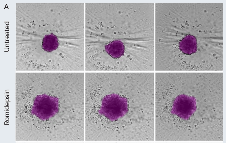
Morphometric analysis of compound treatment on patient-derived spheroids over time. A) Spheroids were monitored using brightfield imaging on days 1, 3, and 5 post-treatment. Images were segmented using SINAP in IN Carta software (magenta overlay).
Machine learning improves image segmentation and objects classification
Automated image analysis is an integral part of most high-content imaging platforms. The ability to monitor cells and organoids in real-time and then extract meaningful information depends on robust image analysis of label-free transmitted light images. Challenges associated with the analysis of brightfield images include low contrast, uneven background, and imaging artifact. A defined set of global parameters would rarely be successful in segmenting objects imaged in brightfield. Recent advances in machine learning improve image analysis workflow and enable more robust image segmentation in complex datasets.
In bioimage analysis, deep learning provides a powerful toolbox to tackle challenging image segmentation and object tracking. Conventional image analysis typically involves defining a fixed set of parameters to segment objects of interest for downstream quantification. However, these predefined parameters do not work for all experiments due to the high variability in experimental conditions. Manual adjustments to the analysis protocol are impractical because of the sheer volume of imaging data in a high-throughput environment.
To overcome these challenges, machine learning tools can be used for image segmentation and object classification to automate the image analysis workflow.
IN Carta Image Analysis Software for machine learning-based, high-throughput analysis
The IN Carta® Image Analysis Software provides an intuitive user interface that includes AI tools in the image analysis workflow. Two key components of IN Carta software that leverage machine learning are the software modules SINAP and Phenoglyphs. Deep-learning-based SINAP enables robust detection of complex objects of interest (e.g., stem cell colonies or organoids) with minimal human intervention to improve accuracy and reliability in the first step of image analysis. The analysis output includes morphological, intensity, and texture measurements.
Classification of data can also be used with machine-learning-based Phenoglyphs. Phenoglyphs takes hundreds of image descriptors extracted by SINAP and creates an optimal set of rules for grouping objects with a similar visual appearance. Both modules utilize unsupervised decisions to generate an initial result that is iteratively optimized through user input. Together, they improve the integrity and accuracy of findings through an easy-to-use, end-to-end workflow.
Apply deep learning to object segmentation with IN Carta SINAP
Automatic object segmentation of microscopy images can be challenging due to the diverse nature of the datasets. IN Carta Image Analysis Software uses SINAP, a trainable segmentation module utilizing a deep convolutional neural network learning algorithm to tackle these challenging issues.
Because SINAP uses deep learning, it can account for significant variability in sample appearance that arises from the test treatments under investigation. By ensuring that each treatment is segmented with an equivalent level of accuracy, the information extracted in this step is reliable and useful to compare treatments in subsequent analysis steps.
Overcome challenges in image segmentation with machine learning-based models:
A) Examples of different biological models are presented that are challenging for quantitative analysis. 3D spheroids grown in microcavity plates produce a shadow around each microcavity that interferes with object segmentation (arrow). 3D organoids are grown in Matrigel, which often produces non-homogenous background due to distortion from the Matrigel dome and objects beyond the imaging planes (box). iPSC grows as relatively flat cultures; as a result, the low contrast (blue arrow) and debris (yellow arrow) hampers robust image segmentation of iPSC colonies.
B) Overview of the model training workflow: Generate images for training > Train model > Test model > Repeat.
C) Main steps to create a model in IN Carta software using SINAP with example images shown. Images are annotated using labeling tools to indicate the objects of interest and background. The annotated image representing ground truth is added to the training set. In the training step, a model is created based on the most-suitable existing model and user-specified annotations. In the example shown, it is necessary to correct the segmentation mask (step 3), repeating steps 1–3.
Apply machine learning to object classification with IN Carta Phenoglyphs
IN Carta Image Analysis Software also includes a trainable object-level classifier module called Phenoglyphs. The Phenoglyphs module uses segmentation measurements to group objects with a similar visual appearance. In doing so, one can assess if a treatment generates a favorable phenotype and can even infer the underlying mechanisms involved. By using machine learning, all visual features can be analyzed simultaneously to optimize the complex set of rules required to assign objects to their correct group. This highly multi-variate and data-driven approach is far more capable of resolving subtle phenotypic differences and is more robust against assigning objects to the incorrect group.
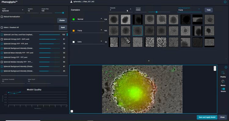
As a user, you only need to review and provide input on a small number of examples for each class before the Phenoglyphs module applies the model to the entire dataset. This approach minimizes the need for user input at the first step of a class assignment, thus saving considerable time.
Machine learning-based applications
Traditional image analysis methods can be incredibly intricate and time-consuming when performed manually or even semi-automatically. There’s always the possibility of human error and bias due to the task’s complex and highly detailed nature. When you add to this the repetitive, lengthy, and often laborious nature of the workflow, there comes the opportunity to apply machine learning.
Learn more about how IN Carta’s deep learning software paired with the ImageXpress Confocal HT.ai system helped remove person-to-person variation, human error, and bias, thereby improving data quality and confidence and optimizing workflow and efficiency.
Export datasets from IN Carta to StratoMineR for deeper insights into your data
IN Carta Image Analysis Software provides robust, quantitative results from complex biological images and datasets utilizing advanced AI technology. Directly import this data into StratoMineR, an intuitive web-based platform which guides users through a typical workflow in analysis of high-content, multi-parametric data.
Latest Resources
Resources for AI, machine learning, and deep learning tools for bio image analysis
Blog
Stem Cell Science and Regenerative Medicine – Technology and Methods Presented at ISSCR 2022
It was another great year at ISSCR 2022! Leaders from across the globe came together to discuss new technologies, share insights, and explore the newest breakthroughs in stem cell…
Publications
AI-Powered Phenotypic Profiling Revolutionizes Drug Discovery
Today, nine out of ten drugs fail in clinical trials. Furthermore, it takes over a decade and an average cost of $2 billion to develop and approve each medicine.1 An important underlyin…
Publications
A Picture Paints a Thousand… New Therapies?
Cell painting is a high-content profiling technology that uses up to six fluorescent dyes to visualize specific cellular components at the single-cell level. The assay essentially “pain…
Scientific Poster
Phenotypic profiling based on the Cell Painting assay
Multiparametric high-content screening approaches, such as the Cell Painting assay, are increasingly being used in many applications ranging from drug discovery programs to functional g…
Scientific Poster
Intestinal organoids for automated screening assays. High content imaging and analysis of organoid morphology
Here we describe a workflow for automation of organoid culture. The automated method utilizes an integrated work-cell, consisting of several instruments providing automated cell culture…
Blog
Life Science and Technology Predictions for 2022
For three decades, Molecular Devices has made scientific breakthroughs possible for academic, pharmaceutical, government, and biotech customers with its innovative life science…
Blog
Engineering Next-gen Organoids with Automated Lab Workflows at #SLAS2022
SLAS2022, the Society for Lab Automation and Screening conference offered another exciting year for learning about innovative laboratory technologies. Whether you attended in-person…
Application Note
Improving the robustness of Cell Painting with a near-infrared label and advanced image and data analytics
Image-based phenotypic profiling approaches such as the widely used Cell Painting assay use high-content imaging along with multiparametric readouts to study biological, genetic, and…
Application Note
Deep Learning-based Image Analysis for Label-free Live Monitoring of iPSC and 3D Organoid Cultures
Complex 3D biological models such as organoids and patient-derived spheroids are gaining popularity in many biomedical research areas because they more closely recapitulate the in vivo…
Blog
Stem cell science insights and breakthroughs presented at #ISSCR2021
If you didn't get a chance to visit us at our poster sessions during ISSCR 2021, don’t fret. We've gathered all our sessions right here for you. The ISSCR Annual Meeting brought…
Scientific Poster
Monitoring organoid development and characterization of calcium oscillation activities in iPSC-derived 3D cerebral organoids
Cerebral organoids are a rapidly developing technology that has great potential for understanding brain development and neuronal diseases. They can also be used for testing effects of c…
Publications
How AI is advancing neurodegenerative disease research
In this guest editorial, explore how artificial intelligence is becoming a powerful tool for drug discovery As part of the SelectScience Advances in Drug Discovery Special Feature, gues…
Blog
Overcome the challenges of high-content cell analysis through AI/machine learning
Artificial intelligence (AI) is finding its way into many aspects of modern life, from autonomous vehicles to voice-powered personal assistants, and even the creation of art. But it…
Blog
How Cell Painting is making its mark on drug discovery
Have you ever heard the old adage, “A picture is worth a thousand words?” When it comes to Cell Painting, this saying is especially true. Cell Painting is a high-content,…
Application Note
High-content phenotypic profiling using the Cell Painting assay
Here, we present a complete workflow for a cell painting assay that can be easily implemented using the ImageXpress Micro system and image analysis with machine learning capabilities
Scientific Poster
AI-based analysis of complex biological phenotypes
Cell-based phenotypic assays have become an increasingly attractive alternative to traditional in vitro and in vivo testing in pharmaceutical drug development and toxicological safety a…
Videos & Webinars

Enhance high-content 3D biology imaging with automated sample preparation

Organoid Innovation Center Walkthrough

An AI-based approach to high-content phenotypic characterization of human iPSC-derived neuronal cells