High-content Imaging
High-content imaging systems ranging from automated digital microscopy to high-throughput confocal imaging systems
What is high-content imaging?
High‑content imaging is an approach to cellular imaging that combines automated microscopy with quantitative image analysis to measure multiple cellular features across large numbers of samples. It is commonly used to analyze cell‑based assays where spatial and phenotypic information is required.
High-content imaging systems from Molecular Devices
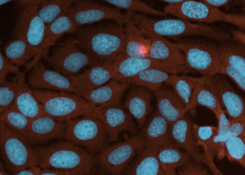
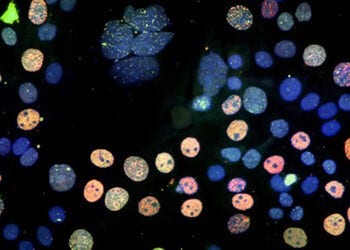
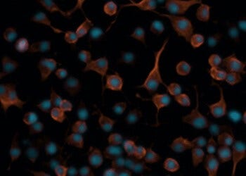
Our ImageXpress® high-content imaging systems range from automated digital microscopy to high-throughput confocal imaging systems. Our state-of-the-art AgileOptix™ technology combines an advanced solid-state light engine, water immersion objectives, a scientific CMOS sensor, and proprietary, dual-spinning disk technology. These systems allow researchers to explore the complex world of cellular biology, capturing high-resolution images of cells and subcellular components with a high level of accuracy and detail.
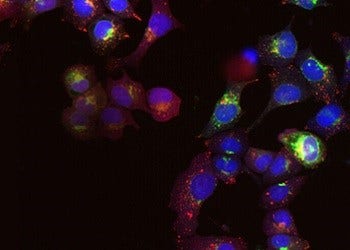
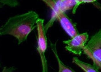
The acquired images are then subjected to in-depth analysis using high-content analysis (HCA) software. This software encompasses powerful algorithms that can extract a wide range of quantitative measurements. With our AI-enabled solutions, researchers can obtain valuable insights into various cellular features, including cell morphology, protein expression levels, subcellular localization, and cellular responses to different treatments or stimuli with increased efficiency.
One key advantage of high-content imaging is its high-throughput capabilities. Researchers can analyze large numbers of cells and images in a relatively short period, enabling them to study complex cellular processes at an unprecedented scale. This feature is particularly useful in drug discovery, toxicology studies, and basic research, as it allows for efficient screening of compounds or investigation of cellular mechanisms.
High-content imaging systems from Molecular Devices
The following systems are part of Molecular Devices’ high‑content imaging portfolio.
FAQs
What is high‑content imaging?
High‑content imaging is an approach to cellular imaging that combines automated microscopy with quantitative image analysis to measure multiple features from cell‑based assays. It is used in experiments where spatial and phenotypic information is derived from images.
How is high‑content imaging different from standard fluorescence microscopy?
High‑content imaging typically involves automated image acquisition and quantitative analysis across many samples, whereas standard fluorescence microscopy is often used for manual or qualitative observation. The distinction is generally described in terms of workflow and data extraction rather than imaging modality alone.
What types of experiments use high‑content imaging?
Examples of experiments that frequently use high‑content imaging include:
1. Phenotypic profiling assays, such as cell painting, where multiple cellular structures are analyzed simultaneously
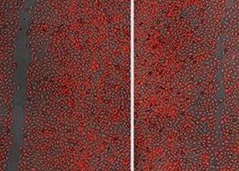
2. Cell migration and invasion assays, which quantify changes in cell movement and morphology over time
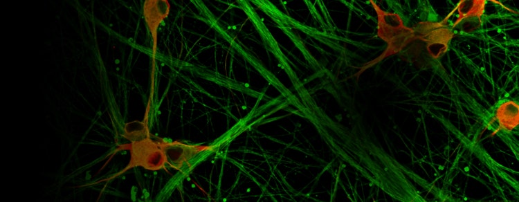
3. Neurite outgrowth assays, used to measure neuronal process extension and branching
4. Drug discovery and screening studies, where cellular responses to compounds are evaluated using image‑based readouts
5. Toxicity screening assays, which assess cellular changes associated with compound‑induced stress or damage
The specific experimental design and measurements depend on the assay, imaging approach, and research objectives.
Latest Resources
Featured cell imaging applications
Cancer Research
Cancer involves changes which enable cells to grow and divide without respect to normal limits, to...
Cell Imaging
Researchers have several options in methods for imaging cells, from phase-contrast microscopy that...

3D Cell Models
Development of more complex, biologically relevant, and predictive cell-based assays for compound..
Live Cell Imaging
Live cell imaging is the study of cellular structure and function in living cells via microscopy....
Cell Counting
Here we discuss the various methods and techniques used to assess proliferation, cytotoxicity, or...
Cell Migration Assays
The movement or migration of cells is often measured in vitro to elucidate the mechanisms of...
Neurite Outgrowth
Neurite outgrowth is assessed by the segmentation and quantification of neuronal processes. These...
Stem Cell Research
Stem cells provide researchers with new opportunities to study targets and pathways that are more...