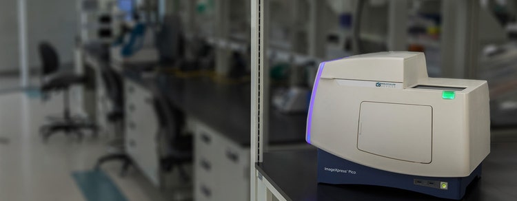
The ImageXpress® Pico Automated Cell Imaging System is more than a digital microscope, combining high-resolution imaging with powerful analysis. Whether running fluorescence imaging or brightfield assays, the automated imager features a comprehensive portfolio of preconfigured protocols for cell-based assays to shorten the learning curve, so you can start running experiments quickly. With features such as Digital Confocal* 2D on-the-fly deconvolution, Autofocus, Live Preview, multi-wavelength cell scoring, and optional IN Carta® Image Analysis Software workflow, the ImageXpress Pico offers you the ability to advance your discoveries in a small, affordable imager.
A simple, easy-to-learn interface for performing quantitative analysis on images acquired from automated microscopy with features such as Digital Confocal 2D real-time deconvolution, Autofocus and Live Preview for advanced region of interest identification. The software enables distributed analysis of images for increased throughput and is ideal for scaling your digital microscopy imaging with slides or microplates. An icon-driven, linear workflow with a range of predefined protocols provides a streamlined user experience.
Powerful analytics combined with an intuitive user interface simplify workflows for image analysis and phenotypic profiling. Advanced features provide the functionality you need to analyze data in 2D, 3D, and 4D - at scale - and deliver real-time insights without the need for complex pre- or post- processing operations. Improve specificity of your image analysis workflows by utilizing the SINAP deep-learning module and see for yourself that Segmentation Is Not A Problem. Put machine learning to work for you and perform complex phenotypic analysis within a user-friendly Phenoglyphs module.
The research environment is constantly changing and today’s scientists require simplified remote access and enhanced laboratory automation. Increased productivity, reduced costs, and consistent performance are all available when combining the ImageXpress® Pico Automated Cell Imaging System with the S-LAB™ plate handler from PAA, and the SCILA incubator from Inheco*.
See the Automated ImageXpress Pico workflow in action
Apoptosis is a process of programmed cell death important to a number of biological processes including embryonic development and normal tissue maintenance. Disruption in the regulation of apoptosis has been implicated in various diseases including cancer. Biochemical events lead to characteristic changes in cell morphology such as cell shrinkage, nuclear fragmentation, chromatin condensation, and mRNA decay, and ultimately cell death. Apoptosis can be initiated via the intrinsic or extrinsic pathways, and both pathways induce cell death by activating caspase enzymes
Cancer researchers need tools that enable them to more easily study the complex and often poorly understood interactions between cancerous cells and their environment, and to identify points of therapeutic intervention. Learn about instrumentation and software that facilitate cancer research using, in many cases, biologically relevant 3D cellular models like spheroids, organoids, and organ-on-a-chip systems that simulate the in vivo environment of a tumor or organ.
Stem cells differentiated into cardiomyocytes are used to screen early for potential toxicological effects of drugs, thus helping to avoid investment in development of drugs which will fail in clinical trials due to cardiac toxicity.
Cell counting is fundamental and critical to numerous biological experiments. Assays such as drug compound toxicity, cell proliferation, and inhibition of cell division have a need to assess the number or density of cells in a well. Automated imaging can greatly speed up the cell counting process while reducing manual labor and human errors. Cells can be counted using a variety of methods, such as label-free cell counting under transmitted light or detection of nuclear dye with fluorescent imaging.
Here we've addressed common applications in infectious disease research including cell line development, binding affinity, viral neutralization, viral titer and more with a focused effort on understanding the SARS-CoV-2 virus in order to develop potential therapies for COVID-19 including vaccines, therapeutics and diagnostics.
The ability of cells to adapt to their environment is mediated by highly controlled signaling pathways. In many cases, the initiation of these pathways by extracellular signals, such as hormones or cytokines, leads to the translocation of proteins from the cytoplasm to the nucleus. Once in the nucleus, these proteins can regulate the expression of target genes. Assays requiring precise identification of sub-cellular structures or molecules within different cellular compartments can benefit from the ability of image deconvolution to improve identification of the specific fluorescence signal against potentially high background caused by out-of-focus light. The ImageXpress Pico system with Digital Confocal 2D on-the-fly deconvolution is capable of measuring the translocation of proteins from the cytoplasm to the nucleus, like NF-κB translocation upon treatment with TNF-α. The enhancement in image resolution and maintenance of quantitative information from the image deconvolution leads to greater statistical significance in the analysis of nuclear translocation.
Live cell imaging is the study of cellular structure and function in living cells via microscopy. It enables the visualization and quantitation of dynamic cellular processes in real time. Live cell imaging encompasses a broad range of topics and biological applications—whether it is performing long-term kinetic assays or fluorescently labeling live cells.
Neurons create connections via extensions of their cellular body called processes. This biological phenomenon is referred to as neurite outgrowth. Understanding the signaling mechanisms driving neurite outgrowth provides valuable insight into neurotoxic responses, compound screening, and for interpreting factors influencing neural regeneration. Using the ImageXpress Micro system in combination with MetaXpress Image Analysis Software automated neurite outgrowth imaging and analysis is possible for slide or microplate-based cellular assays.
Pluripotent stem cells can be used for studies in developmental biology or differentiated as a source for organ-specific cells and used for live or fixed cell-based assays on slides or in multi-well plates. The ImageXpress system has utility in all parts of the stem cell researcher’s workflow, from tracking differentiation, to quality control, to measuring functionality of specific cell types.
Toxicology is the study of adverse effects of natural or man-made chemicals on living organism. It is a growing concern in our world today as we are exposed to more and more chemicals, both in our environment and in the products we use.
Customer Breakthrough
The production of cow leather consumes vast quantities of energy and water and contributes to climate change via the emission of methane gas. To reduce the environmental impact of le…
Blog
Target discovery and drug development rely heavily on 2D cell and animal models to decipher efficacy and toxic effect of drug candidates. Yet, 90% of candidates fail to make it past…
News
SAN JOSE, Calif., and UTRECHT, Netherlands, Feb. 13, 2023 – Molecular Devices, LLC., a leading provider of high-performance life science solutions, and HUB Organoids (HUB), the pioneer…
Blog
As we enter the era of sophisticated drug discovery with gene therapy and personalized medicine, we need to be prepared to study complex diseases, assess the therapeutic effect of…
Publications
More researchers are using gene editing to build disease models that better represent human tissues' complex biology, signaling a shift away from 2D cell culture or animal models to…
Publications
3DPrint.com, the leading source for actionable intelligence related to 3D printing technology and the larger AM industry, covers our collaboration…
Publications
Following our acquisition of Cellesce Ltd, Tanya Samazan from Instrument Business Outlook dove deep into conversation with Molecular Devices President Susan Murphy and Cellesce CEO V…
News
Turnkey platform integrates flexible robotic automation with high-content imaging of complex 3D cellular models, enabling high-volume organoid screening SAN JOSE, Calif., Jan. 4, 2023…
Application Note
3D bioprinting is defined as the additive deposition of cells and biocompatible materials to build biologically functional 3D structure or artificial tissue models
News
First-of-its-kind technology from Cellesce creates consistent patient-derived organoids for large scale drug screening Acquisition strengthens Molecular Devices’ position as a 3D…
Publications
Gone are the days of only measuring single parameters in cell-based experiments. Instead, researchers should widen their focus, namely with the help of innovations like the Cell Pain…
Publications
We recently partnered with HeartBeat.bio to automate and scale the production of cardioids — 3D cell models of the heart that can more accurately recapitulate human biology.
…
Publications
In April 2021, we launched our Organoid Innovation Centre (OIC), an initiative designed to help scientists leverage the potential of 3D biology throughout the drug discovery process.…
Application Note
The intestinal crypt system is an important part of the intestinal organoid where mature organoids have more complex and numerous crypt structures. Yet, due to their high turnover rate…
Application Note
3D cellular models are increasingly popular in many areas of research because they recapitulate the cellular 3D microenvironment better than cells grown in 2D monolayer.
Scientific Poster
3D cell culture as a model system is increasingly popular because it recapitulates the in vivo microenvironment better than 2D cell cultures. Organoids have the capacity for stable d…
Blog
From customer feedback to workflow improvements The path to understanding complex biological processes and diseases is paved with a lot of challenges. As the desired level of…
Blog
If you didn't get a chance to visit us at our poster sessions during ISSCR 2021, don’t fret. We've gathered all our sessions right here for you. The ISSCR Annual Meeting brought…
Brochure
The CellReporterXpress® Automated Image Acquisition and Analysis Software works with the ImageXpress® Pico Automated Cell Imaging System. It has a clean, easy-to-learn interface for…
eBook
From endpoint and live-cell imaging applications to imaging and analyzing 2D monolayers, 3D cell models, and organ-on-a-chip platforms, the world of cellular imaging is very diverse.…
Publications
Lab Manager speaks to Dan O’Connor, vice president, drug discovery, Molecular Devices, about the company’s Organoid Innovation Center in San Jose, CA. The center is 180 sq. ft., with…
Scientific Poster
Our first-in-class automated 7-factor neuronal profiling assay enables comprehensive quantitative description of neuronal health. NeuroHTS™ maximizes the capacity of imaging-type of…
Flyer
Environmental control mimics the cell environment and enables you to run multi-day studies, time-lapse, and live cell assays. With CellReporterXpress™ software, the ImageXpress® Pico…
Blog
There have been significant advancements in microscopy and camera technology, as well as advancements in technologies for labeling molecules of interest over the past decade. These…
Publications
Molecular Devices, a provider of innovative life science technology, recently unveiled a brand new, first-of-its-kind Organoid Innovation Center. Situated at the company’s global hea…
Application Note
From studying disease to evaluating drug and environmental compound-induced toxicity, cell-based assays aimed at monitoring cellular health often focus on mitochondria.
Brochure
The ImageXpress® Pico system does more than imaging—it offers unparalleled analysis capabilities that simplifies image analysis for cell-based assays.
Blog
Cheryl Bell talks about making the transition from manual microscopy to automated microscopy Our Field Applications Scientist, Cheryl Bell, was recently featured in a Women in…
Scientific Poster
There is an increasing interest in using three-dimensional (3D) cell structures for modeling tumors, organs, and tissue to accelerate translation research. Significant progress has b…
Application Note
The formation of new blood vessels from existing ones, angiogenesis, is a critical physiological process involved in vertebrate development and wound healing. The process of…
eBook
Gain insights and expedite studies for 2D and 3D cellular structures using automated cellular imaging.
Blog
For over 40 years, Molecular Devices has been at the forefront of technological advances which have contributed to significant scientific breakthroughs. To kick off the new year, we…
Blog
Kayla Hill discusses the latest trends in cellular imaging We recently hosted a webinar with our Field Applications Scientist, Kayla Hill, PhD, who explored high-content analysis…
Application Note
There is a growing trend to develop screening assays using three-dimensional (3D) cell culture.
Flyer
Molecular Devices provides full-service, customization, and automation to meet your research needs. Our team includes scientists and engineers who can customize our instruments, as w…
Flyer
Designed for the ImageXpress® Pico Automated Cell Imaging System, and utilizing CellReporterXpress® Image Acquisition and Analysis Software, the Digital Confocal* Option provides 2D…
Customer Breakthrough
Understand the binding characteristics of mast cells and hyaluronan in the extracellular matrix of RSV-infected human lung fibroblasts
Respiratory sy…
Publications
With each passing day, continuous advancements are being made in the studies that cutting-edge instruments can perform and in the complexity of the biological samples that are being…
Publications
OUR ABILITY to image cells has come a long way since the pioneering days of Galileo Galilei and Antonie van Leeuwenhoek. The multiple imaging techniques available today range from si…
Customer Breakthrough
The Tomlinson lab, in the School of Molecular and Cellular Biology at the University of Leeds, UK, investigates protein funct…
Application Note
Since the discovery of green fluorescent protein in the 1960s, a wide variety of fluorescent proteins (FP) and variants have been developed to span the entire visible light spectrum.…
Publications
Depending on your application and exactly what you are researching, there are multiple imaging techniques available, ranging from confocal microscopy to electron microscopy and more…
Application Note
Widefield microscopy is a powerful tool that has been utilized to investigate cellular and molecular processes involved in cell signaling.
Application Note
The study of apoptosis is a critical aspect of drug discovery and development. Additionally, studying the relationship between apoptosis and other factors, such as oxidative stress, is…
Publications
Ever since the discovery that tumors undergo angiogenesis in order to grow and survive, researchers have sought to better understand the detailed mechanism of new blood vessel format…
Scientific Poster
There is an increased need for expanding variety and complexity of cell-based biologic research and drug discovery.
Publications
Angiogenesis is an important field of research and a focus for cancer therapeutics. In this interview, Dr. Bas Trietsch, CTO, MIMETAS, introduces a new solution for the study of angi…
Application Note
There is an increased need to expand the variety and complexity of cell-based assays for biological research and drug discovery. Live-cell assays allow monitoring of cell responses in…
Application Note
Macrophages originate from blood monocytes that leave the circulation and penetrate various tissues where they differentiate into macrophages.
Application Note
The ability to accurately quantitate cell number in multi-well microplates enables a multitude of biological applications that study cell health or proliferation.
Scientific Poster
Monitor cell responses in real time to acquire important qunatitative insights
Scientific Poster
Screen models that mimic in vivo tumor microenvironment using novel 3D cell culture platform, VersaGel and Symphony
Scientific Poster
Effectively image and analyze cell migration using a scratch assay or a dissolvable biocompatible gel
Application Note
Cell migration is an essential process required for many biological events including embryonic development, wound healing, cancer metastasis, and immunological responses.
eBook
Gain valuable insights into neurobiology, from evaluation of ion channels to analysis of 3D neuron organoids.
Scientific Poster
Develop functional and morphological readouts for testing effects of different compounds in a multi-parametric assay format
Scientific Poster
Effectively image and analyze cell migration using a dissolvable biocompatible gel.
Flyer
These assay kits are a family of fluorescence-based reagents for the assessment of cell viability, cell proliferation, and apoptosis events using mammalian cells. The kits employ a n…
eBook
This new eBook discusses early drug safety testing using HTS iPSC-derived cells with imaging. Download it now!
Application Note
Evaluate compound-specific effects on neurite outgrowth using automated imaging
Application Note
Detect and quantify compound effects on the process of autophagy on the ImageXpress Pico system. The PC12 human neuroblastoma cell line was used as a model for assay development.
Scientific Poster
Automate complex cell-based assays with multi-parametric readouts while maintaining high data quality and precision.
Publications
Developments in informatics solutions and high-content imaging instruments, increased focus on cell-based research, and rising government support of research and development for drug…
Customer Breakthrough
MIMETAS offers the OrganoPlate®, a unique 3D organ-on-a-chip platform. The OrganoPlate® is a fully compatible microfluidic culture plate, enabling testing of compounds in any through…
Scientific Poster
There is a great need to automate complex cell-based assays with multi-parametric read-outs while maintaining high data quality and precision. A compact, automated imaging system was…
Application Note
Automated cellular imaging is an efficient method for analyzing compound effects on cell phenotypes including morphology, viability, and marker expression.
Application Note
Mitochondrial function, a key indicator of cell health, can be assessed by monitoring changes in mitochondrial membrane potential (MMP)1,2.
Application Note
Live/dead assays are utilized in a wide variety of research applications including investigation of cytotoxic effects of various compounds, treatments, or changes in gene expression.
Application Note
Apoptosis is a process of programmed cell death that occurs in multicellular organisms.1,2 Biochemical events lead to characteristic changes in cell morphology and cell death.
Brochure
Learn about our imaging family and which solution is best suited to answer your biological questions.
Flyer
Replace tedious manual microscope manipulations with the fully automated ImageXpress® Pico Automated Cell Imaging System. Simply place samples into the system and follow the icon-dri…
Application Note
The discovery and development of novel drugs is a difficult and expensive process due to lack of tools and techniques to accurately reproduce the human physiological response in the…
Brochure
Molecular Devices is a leading provider of high-performance bioanalytical measurement solutions for life science research, pharmaceutical development, and biotherapeutic discovery. O…
Ananda Neurite Untangled
Enhancing 3D Disease Models: Automated, High-Throughput, Phenotypic Screening with Organ-on-a-Chip
3D Imaging Seminar Series: Fast and simple 3D spheroid analysis using automated high-content imaging
Automated Cell Imaging with the ImageXpress Pico system
Immunology and Vaccine Development Workflow
Hybridoma Workflow
How to install the ImageXpress Pico Environmental Control System
Accelerate your screening with high-content and automated imaging
Accelerating study of viral infection and therapeutics with microplate-based detection and high-throughput screening
ImageXpress Pico Automated Imaging System Virtual Tour
Magnetic 3D Bioprinting, 3D Cell Culture in a 2D Workflow
Developing high-throughput organ-on-a-chip tissue models for drug discovery using high-content imaging
Automated Imaging and You – Quantitative microscopy for every lab, Powerful data for all
How to easily image slides and regions of interest with Live Preview on the ImageXpress Pico
Standard Widefield Imaging
See what’s new with CellReporterXpress
A Product Manager's Tour of the ImageXpress Pico
Configuring environmental control settings on the ImageXpress Pico
How to perform a z-stack image acquisition using CellReporterXpress
Cellular images to results in minutes with automated imaging
Setting up acquisition and analysis on the ImageXpress Pico
Transmitted light cell scoring on the ImageXpress Pico
Reviewing completed experiments on the ImageXpress Pico
ImageXpress Pico Automated Cell Imaging System-Interactive Demo
ImageXpress Pico Automated Cell Imaging System
Latest Citations: For a complete list, please click here .
*Source: https://scholar.google.com/
Responses to therapy often cannot be exclusively predicted by molecular markers, thus evidencing a critical need to develop tools for better patient selection based on relations between tumor phenotype and genotype.
Rett syndrome (RTT) is a neurodevelopmental disorder caused by pathogenic variants leading to functional impairment of the MeCP2 protein.
Personalized medicine holds tremendous promise for improving safety and efficacy of drug therapies by optimizing treatment regimens. Rapidly developed patient-derived xenografts (pdx) could be a helpful tool for analyzing the effect of drugs against an individual’s tumor by growing the tumor in an immunodeficient animal. Severe combined immunodeficiency (SCID) mice enable efficient in vivo expansion of vital tumor cells and generation of personalized xenografts. However, they are not amenable to large-scale rapid screening, which is critical in identifying new compounds from large compound libraries. The development of a zebrafish model suitable for pdx could facilitate large-scale screening of drugs targeted against specific malignancies. Here, we describe a novel strategy for establishing a zebrafish model for drug testing in leukemia xenografts. We used chronic myelogenous leukemia and acute myeloid leukemia for xenotransplantation into SCID zebrafish to evaluate drug screening protocols. We showed the in vivo efficacy of the ABL inhibitor imatinib, MEK inhibitor U0126, cytarabine, azacitidine and arsenic trioxide. We performed corresponding in vitro studies, demonstrating that combination of MEK- and FLT3-inhibitors exhibit an enhanced effect in vitro. We further evaluated the feasibility of zebrafish for transplantation of primary human hematopoietic cells that can survive at 15 day-post-fertilization. Our results provide critical insights to guide development of high-throughput platforms for evaluating leukemia.
Mouse brain contains over 100 million neuronal, glial, and other support cells. Developing neurons and astrocytes synthesize their own cholesterol, and disruption of this process can occur by both genetic and chemical mechanisms. In this study we have exposed cultured murine neurons and astrocytes to six different prescription medications that cross the placenta and blood–brain barriers and analyzed the effects of these drugs on cholesterol biosynthesis by an LC–MS/MS protocol that assays 14 sterols and 7 oxysterols in a single run. Three antipsychotics (haloperidol, cariprazine, aripiprazole), two antidepressants (trazodone and sertraline), and an antiarhythmic (amiodarone) inhibited one or more sterol synthesis enzymes. The result of the exposures was a dose-dependent increase in levels of various sterol intermediates and a decreased level of cholesterol in the cultured cells. Four prescription medications (haloperidol, aripiprazole, cariprazine, and trazodone) acted primarily on the DHCR7 enzyme. The result of this exposure was an increase in 7-dehydrocholesterol in neurons and astrocytes to levels that were comparable to those found in cultured neurons and astrocytes from transgenic mice that carried a Dhcr7 pathogenic mutation modeling the neurodevelopmental disorder Smith–Lemli–Opitz syndrome.
Human lung fibroblasts (HLFs) treated with the viral mimetic polyinosine-polycytidylic acid (poly I:C) form an extracellular matrix (ECM) enriched in hyaluronan (HA) that avidly binds monocytes and lymphocytes. Mast cells are important innate immune cells in both asthma and acute respiratory infections including respiratory syncytial virus (RSV); however, the effect of RSV on HA dependent mast cell adhesion and/or function is unknown. To determine if RSV infection of HLFs leads to the formation of a HA-enriched ECM that binds and enhances mast cell activity primary HLFs were infected with RSV for 48 h prior to leukocyte binding studies using a fluorescently labeled human mast cell line (LUVA). Parallel HLFs were harvested for characterization of HA production by ELISA and size exclusion chromatography. In separate experiments, HLFs were infected as above for 48 h prior to adding LUVA cells to HLF wells. Co-cultures were incubated for 48 h at which point media and cell pellets were collected for analysis. The role of the hyaladherin tumor necrosis factor-stimulated gene 6 (TSG-6) was also assessed using siRNA knockdown. RSV infection of primary HLFs for 48 h enhanced HA-dependent LUVA binding assessed by quantitative fluorescent microscopy. This coincided with increased HLF HA synthase (HAS) 2 and HAS3 expression and decreased hyaluronidase (HYAL) 2 expression leading to increased HA accumulation in the HLF cell layer and the presence of larger HA fragments. Separately, LUVAs co-cultured with RSV-infected HLFs for 48 h displayed enhanced production of the mast cell proteases, chymase, and tryptase. Pre-treatment with the HA inhibitor 4-methylumbelliferone (4-MU) and neutralizing antibodies to CD44 (HA receptor) decreased mast cell protease expression in co-cultured LUVAs implicating a direct role for HA. TSG-6 expression was increased over the 48-h infection. Inhibition of HLF TSG-6 expression by siRNA knockdown led to decreased LUVA binding suggesting an important role for this hyaladherin for LUVA adhesion in the setting of RSV infection. In summary, RSV infection of HLFs contributes to inflammation via HA-dependent mechanisms that enhance mast cell binding as well as mast cell protease expression via direct interactions with the ECM.