3D imaging and analysis of cell models like spheroids, organoids, and organ-on-a-chip biology for screening cancer therapeutics
Cancer involves changes which enable cells to grow and divide without respect to normal limits, to invade and destroy adjacent tissues, and ultimately to metastasize to distant sites in the body. Cancer researchers need tools that enable them to more easily study the complex and often poorly understood interactions between cancerous cells and their environment, and to identify points of therapeutic intervention.
Learn about our high-content imaging systems and analysis software solution that facilitate cancer research using biologically relevant 3D cellular models like spheroids, organoids, and organ-on-a-chip systems that simulate the in vivo environment of a tumor or organ.

Figure 1: A workflow for testing spheroids in a high-throughput screening environment. A single spheroid can be grown in a 96- or 384-well plate, treated with compound, and stained with a cocktail of dyes that can be imaged without washing. Spheroids may also be fixed if desired. (Right) Transmitted light images of HCT116 cells were taken over the course of 63 hours using Timelapse acquisition on ImageXpress Micro confocal System to show the formation of a spheroid (10X objective).
Advantages of 3D imaging technology for cancer spheroids
Cancer spheroids mimic tumor behavior far more effectively than standard 2D cell cultures. Such 3D spheroid models are being successfully used in screening environments for identifying potential cancer therapeutics. These culture systems can be used in multiparametric analysis to quantify different biological outputs, accelerating cancer drug development.
Key benefits include:
- The development of 3D high-content imaging signifies a major step in facilitating more relevant and accurate testing
- 3D culture systems can rapidly produce uniform human cancer cell spheroids that can be used in high-throughput format to accelerate cancer drug development
- Research with confocal 3D image analysis of cancer cells has enabled multiparametric characterization of many biological outputs
Workflow for analyzing 3D cancer spheroids in high-throughput screening environment
Spheroids can be grown in 96- or 384-well plates, treated with compounds, and stained with dyes that reveal the cellular processes and pathways at work. In some cases, spheroids can be imaged without washing; they may also be fixed if desired.
Simplify your oncology workflow with a broad range of imaging, cellular screening, and microplate reader systems.
The workflow illustrates a simplified process for analyzing spheroids and highlights systems to help you streamline research and increase your throughput.
-
Culture spheroids – Cancer cells can be cultured directly in an ultra-low attachment (ULA), round bottom plate, or other labware to develop the typical morphology of a spheroid. Other labware allows one to grow multiple spheroids in a single well.
-
Treat with compounds – After spheroid formation, compounds at the desired concentrations are added into the wells, and then incubated for one to several days, depending on the mechanism being studied.
-
Stain for markers – After compound treatment is complete, stains are added directly to the media. Stains that require no washing can be used to avoid disturbing spheroids, but spheroids can be carefully washed, even using automation, if necessary.
-
Acquire spheroid images – Images within the body of the spheroid can be captured individually or as a z-stack (multiple images taken at differing depths) using specialized imaging equipment.
-
Analyze cancer cells – Use cellular imaging analysis software to run quantitative analysis of the cell images to monitor the expression of different markers and to quantify biological readouts.
High-throughput confocal imaging of spheroids for screening cancer therapeutics
In recent years, there has been significant progress in development of in vitro aggregates of tumor cells for use as models for in vivo tissue environments. When seeded into a well of a low-attachment round bottom microplate, these aggregates will form a discrete spheroid. Spheroids are believed to mimic tumor behavior more effectively than regular two dimensional (2D) cell cultures because, much like tumors, they contain both surface-exposed and deeply buried cells, proliferating and non-proliferating cells, and a hypoxic center with a well-oxygenated outer layer of cells. Such 3D spheroid models are being successfully used in screening environments for identifying potential cancer therapeutics.
While there are several challenges to developing robust spheroid assays, using automated high-throughput, high-content imaging is a significant step in facilitating more relevant testing of chemotherapeutic drug candidates
- Capture an entire spheroid in one field-of-view at 20X magnification
- Screen biologically relevant 3D spheroids in 96- or 384-well format
- Use confocal imaging to accurately detect cellular responses
- Conserve storage space by saving only 2D reconstructions of the z plane images
Figure 1. Rapidly screen 3D spheroids in microplates
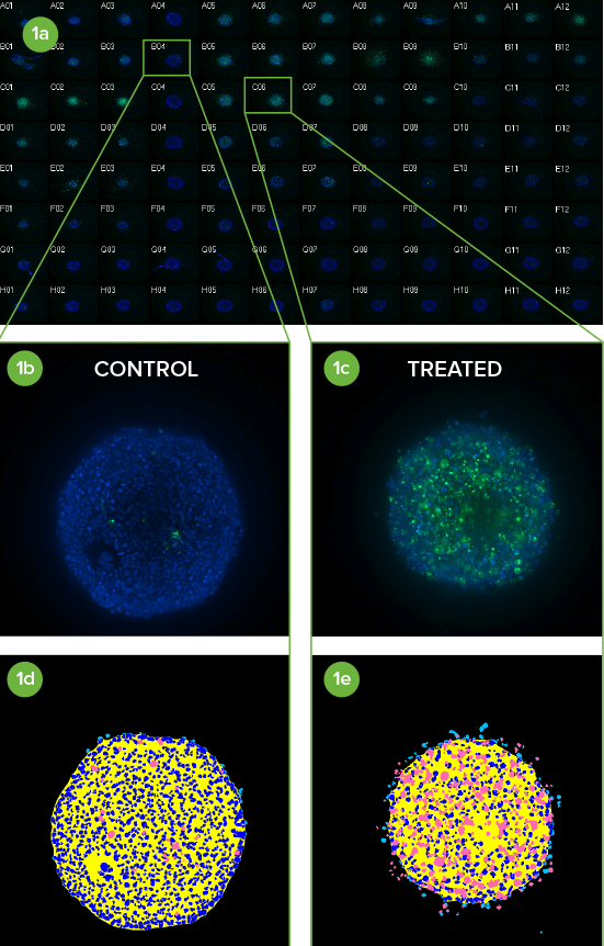
(1a) Montage of image thumbnails of HCT116 spheroids in a 96-well plate treated with compounds and imaged with a 10X Plan Fluor objective. Hoechst stained nuclei (blue) are overlaid with CellEvent Caspase 3/7 apoptosis marker (green).
(1b) Untreated controls are in column 4 and (1c) a Caspase 3/7 response is evident in columns 5–7 where Paclitaxel was serially diluted 1:3 from 1 µM in Row A (replicates of 3 across).
(1d, 1e) Eleven z planes were combined into a 2D Maximum Projection image and analyzed with a simple custom module. Raw images showing low and high degree of apoptosis with their corresponding segmentation masks are shown (royal blue = nuclei, pink = apoptotic cells).
Applications and assays
Molecular Devices, an industry leader in cellular imaging, provides a wide range of tools to support life science research, drug discovery, and high-throughput screening. Our high-content imaging systems can drive the success of your bioanalytical cancer research efforts. We also provide several configurations of our multi-mode microplate readers as well as a line of easy-to-use microarray scanners.
Learn more about how our technology can help your research in cancer therapeutics.
-
3D cancer cell spheroids
The rapid progress of cancer research over the past few decades has seen the rise of spheroids—3D aggregates of cells in culture—as a valuable research tool that provides more physiological relevance than traditional 2D cell culture.
Learn how spheroids formed using a variety of cancer cell types are analyzed using methods featured in our application notes.
- Acquire and analyze images of FUCCI spheroids
- 3D analysis and morphometric characterization of compound effects on cancer spheroid cultures
- High-throughput confocal imaging of spheroids for screening cancer therapeutics
- Multi-parameter imaging assay for measuring toxicity in a tumor model
- 3D Imaging of cancer cell spheroids
- High-throughput screening of 3D cell cultures with multiple high density scaffold-free spheroids for cancer toxicity studies
Angiogenesis
Angiogenesis is an important factor in blood vessel formation and cancer progression. in vitro with tube formation assays is an established method to evaluate angiogenesis. Recent newly developed methods like organ-on-chip models or in vivo using zebrafish as a model system provide different insights in studying angiogenesis. Importantly, scaling up these assays for screening poses significant challenges for image acquisition and analysis.
Learn how our ImageXpress high-content imaging system and MetaXpress software provide a streamlined workflow for the assessment of angiogenesis using models such as zebrafish, tube formation assays, and organ-on-a-chip.
-
Autophagy, DNA damage
Discovery and evaluation of anti-cancer therapies include development of cell-based models, screening for novel drugs, and understanding the relevant mechanisms of action. The cellular effects of processes like DNA damage and autophagy, a regulated process of degrading and recycling damaged proteins and organelles in response to cellular stress, can be analyzed effeciently using automated cell imaging.
Learn how automated cellular imaging provides an effecient method for analyzing the cellular effects of anti-cancer compounds.
Cell migration
Cell migration is broadly defined as the movement of cells from one location to another. It is an essential process required for many biological events including embryonic development, wound healing, and immunological responses. The invasion of tumor cells into surrounding tissues, as well as metastasis, are areas of cancer that can be studied using in vitro cell migration methods.
Learn how to measure cellular migration over time and perform real-time analysis.
-
Disease Modeling
Disease modeling is the process of using stem cells to derive 3D cell or tissue models that display the key pathological processes taking place in the actual disease. Disease modeling is vital to our understanding of how diseases progress and developing more effective therapies without the need for donor tissues.
Find out about cell-based and automated organoid assays for disease modeling, 3D imaging, and the role of disease modeling in immuno-oncology and neurodegenerative disease studies.
- Webinar: Establishing and imaging 3D oncology models
- Webinar: Disease modeling in the 21st century: Automated organoid assays with 3D imaging
- Publication: Disease Modeling with 3D Cell-Based Assays Using a Novel Flowchip System and High-Content Imaging
- Webinar: Capturing the complexity of 3D biology: Organoids for disease modelling and toxicity research
- Poster: Novel assay methods for cancer patient derived organoids
- Customer Breakthrough: Bioneer use the ImageXpress Micro Confocal for high-throughput imaging of 3D disease models
- Webinar: High-throughput, organoid-derived organ-on-a-chip systems for drug discovery and disease modelling
Live cell imaging
Live cell imaging is the study of cellular structure and function in living cells via microscopy. It enables the visualization and quantitation of dynamic cellular processes in real time.
Live cell imaging encompasses a broad range of biological applications, from long-term kinetic assays to fluorescently labeling live cells.
Learn how cellular processes are analyzed using methods featured in our application notes
-
Luminescence-based methods
Cancer studies often utilize luminescence-based methods for measuring parameters such as cell viability in response to drug treatment, and biomolecular interactions occurring in response to stimulation of cell signaling pathways.
Here, we describe how the SpectraMax® iD5 Multi-Mode Microplate Reader yields optimal results for luminescence assays. Find out how you can:
Organoid technology / Organ-on-a-Chip
Organoid technology, like organ-on-a-chip emulates organ physiology though co-culture of cells in a supportive 3D matrix and use of microfluidic channels to perfuse nutrients or compounds over the resulting cellular structures. It is rapidly gaining popularity as a biologically relevant screening model for new drugs and toxicity.
- 3D image analysis and characterization of angiogenesis in organ-on-a-chip model
- Video: Physiologically-Relevant Tissue Models Using a High-Throughput Organ-on-a-Chip Platform
- High-content assay for morphological characterization of 3D neuronal networks in a microfluidic platform
- Webinar: Developing high-throughput organ-on-a-chip tissue models for drug discovery using high-content imaging
- Poster: Water immersion objectives for automated high-content imaging to improve precision and quality of complex biological assays
-
Organoids
Organoids are three-dimensional (3D) multi-cellular microtissues that are designed to closely mimic the complex structure and functionality of human organs. Organoids typically consist of a co-culture of cells which demonstrate a high order of self-assembly to allow for an even better representation of complex in vivo cell responses and interactions, as compared to traditional 2D cell cultures.
Tumoroids
A tumoroid is a special type of organoid derived from primary tumor cells harvested from cancer patients. Because tumoroids can perfectly mimic a typical tumor microenvironment (TME), they give us insights into the impact of TME on tumor progression. That’s why tumoroids boast massive potential for accurate and cost-effective anticancer drug discovery.
Learn how our ImageXpress High-Content Imaging System can help you visualize tumoroids during drug screening:
- Webinar: Establishing and imaging 3D oncology models
- Webinar: Disease modeling in the 21st century: Automated organoid assays with 3D imaging
- Publication: Disease Modeling with 3D Cell-Based Assays Using a Novel Flowchip System and High-Content Imaging
- Webinar: Capturing the complexity of 3D biology: Organoids for disease modelling and toxicity research
- Poster: Novel assay methods for cancer patient derived organoids
Resources to aid in your cancer research
Publications
Benefits of Real-Time Live-Cell Imaging
Three-dimensional (3D) cell-based assays replace a host of tissue, cell, and animal test models but have a reputation for lacking time resolution. For example, traditional, endpoint cell-bas…
Application Note
Label-free cell segmentation with IN Carta SINAP application module
Label-free cell analysis provides a better alternative to using fluorescent dyes as it enables scientists to image live cells under near-native conditions – examining biological processes…
Blog
Engineering Next-gen Organoids with Automated Lab Workflows at #SLAS2022
SLAS2022, the Society for Lab Automation and Screening conference offered another exciting year for learning about innovative laboratory technologies. Whether you attended in-person…
Application Note
Improving the robustness of Cell Painting with a near-infrared label and advanced image and data analytics
Image-based phenotypic profiling approaches such as the widely used Cell Painting assay use high-content imaging along with multiparametric readouts to study biological, genetic, and…
Blog
Enabling 3D High-Content Imaging and Analysis on the Organ-on-a-Chip Platform
Imagine having the ability to mimic the human biological environment for disease modeling and drug screening and doing so in a micro-scale system. With the development of organ-on-a-…
Scientific Poster
Automation and high content imaging of 3D triple-negative breast cancer patient-derived
Triple negative breast cancer is a clinically aggressive tumor subtype, with high rates of metastasis, recurrence, and drug resistance. Currently there are no clinically approved small molec…
Scientific Poster
Improving the robustness of Cell Painting with a near-infrared label and advanced image and data analytics
Image-based phenotypic profiling approaches, such as the widely-used Cell Painting assay, use high-content imaging along with multiparametric readouts to study biological, genetic, and chemi…
Publications
Talking Techniques | Organoids: advancing drug discovery and cancer research
"As the transition from 2D to 3D cell cultures, or organoids, as the gold standard for modeling basic biology and disease continues, these models are being utilized in ever more intricate an…
Blog
Stem cell science insights and breakthroughs presented at #ISSCR2021
If you didn't get a chance to visit us at our poster sessions during ISSCR 2021, don’t fret. We've gathered all our sessions right here for you. The ISSCR Annual Meeting brought…
Scientific Poster
Organoids for disease modeling and in vitro drug screening
3D cell models representing various tissues were successfully used for studying complex biological effects, tissue architecture, and functionality. However, the complexity of 3D models remai…
Scientific Poster
Monitoring organoid development and characterization of calcium oscillation activities in iPSC-derived 3D cerebral organoids
Cerebral organoids are a rapidly developing technology that has great potential for understanding brain development and neuronal diseases. They can also be used for testing effects of compou…
Scientific Poster
High-throughput assessment of compound-induced pro-arrhythmic effects in human IPSC-derived cardiomyocytes
Development of biologically relevant and predictive cell-based assays for compound screening and toxicity assessment is a major challenge in drug discovery. The focus of this study was to es…
Scientific Poster
Deep learning-based image analysis for label-free live monitoring of iPSC 3D organoid cultures
Complex 3D biological models such as organoids and patient derived spheroids are gaining popularity in many biomedical research areas because they more closely recapitulate the in vivo tissu…
Publications
Project Profile: Organoid Innovation Center
Lab Manager speaks to Dan O’Connor, vice president, drug discovery, Molecular Devices, about the company’s Organoid Innovation Center in San Jose, CA. The center is 180 sq. ft., within a lar…
Blog
Tips for running a successful live cell imaging experiment
There have been significant advancements in microscopy and camera technology, as well as advancements in technologies for labeling molecules of interest over the past decade. These…
Publications
Shaping the Future of Organoid Research
Molecular Devices, a provider of innovative life science technology, recently unveiled a brand new, first-of-its-kind Organoid Innovation Center. Situated at the company’s global headquarter…
Blog
Overcome the challenges of high-content cell analysis through AI/machine learning
Artificial intelligence (AI) is finding its way into many aspects of modern life, from autonomous vehicles to voice-powered personal assistants, and even the creation of art. But it…
Publications
Using 3D cancer cell models to push forward personalized medicine
Using 3D cancer cell models to push forward personalized medicine: an interview with Angeline Lim, PhD, of Molecular Devices Editor-in-Chief, Francesca Lake, speaks to Angeline Lim of Mole…
Publications
Disease Modeling with 3D Cell-Based Assays Using a Novel Flowchip System and High-Content Imaging
There is an increasing interest in using three-dimensional (3D) cell structures for modeling tumors, organs, and tissue to accelerate translational research. We describe here a novel automat…
Scientific Poster
Novel assay methods for cancer patient derived organoids
In recent years, researchers have transitioned from traditional 2D assays to more complex 3D cell models, as they are shown to recapitulate the in vivo environment and serve as a more predic…
Blog
How Cell Painting is making its mark on drug discovery
Have you ever heard the old adage, “A picture is worth a thousand words?” When it comes to Cell Painting, this saying is especially true. Cell Painting is a high-content,…
eBook
Cellular Imaging Insights
Gain insights and expedite studies for 2D and 3D cellular structures using automated cellular imaging.
Application Note
High-throughput screening of 3D cell cultures with multiple high density scaffold-free spheroids for cancer toxicity studies
3D spheroid models for cancer research are gaining popularity because they better mimic the in vivo tissue architecture, gene expression and metabolic profile of tumors compared to…
Scientific Poster
AI-based analysis of complex biological phenotypes
Cell-based phenotypic assays have become an increasingly attractive alternative to traditional in vitro and in vivo testing in pharmaceutical drug development and toxicological safety assess…
Publications
The workflow game-changer: combined technologies for efficiency
Microfluidic flowchip technology combined with three-dimensional (3D) imaging is a powerful duo poised to change the future of drug discovery and development. The integration of combined tec…
Application Note
3D image analysis and characterization of angiogenesis in organ-on-a-chip model
Angiogenesis is the physiological process of formation and remodeling of new blood vessels and capillaries from pre-existing blood vessels. This can be achieved through endothelial…
Publications
3D Microscopy Keeps Getting Faster, Smarter, Leaner
With each passing day, continuous advancements are being made in the studies that cutting-edge instruments can perform and in the complexity of the biological samples that are being examined…
Publications
From 3D high throughput to simple automated imaging: Cellular imaging within reach of every laboratory
OUR ABILITY to image cells has come a long way since the pioneering days of Galileo Galilei and Antonie van Leeuwenhoek. The multiple imaging techniques available today range from simple lig…
Publications
What 3D imaging means for cancer drug discovery
Finding a cure for cancer has been called this century’s “moonshot.” As a senior scientist at Molecular Devices, I know first-hand that accelerating cancer drug discovery is a priority of no…
Publications
Revolutionizing early drug discovery for immuno-oncology and neurodegenerative disease modeling
The development of 3D cellular models of disease has enabled researchers to better recapitulate in vivo cell environments. As 3D models become more prevalent in the drug discovery industry,…
Publications
Everything you need to know about the ImageXpress Pico Automated Cell Imaging System
Depending on your application and exactly what you are researching, there are multiple imaging techniques available, ranging from confocal microscopy to electron microscopy and more - each h…
Publications
LabTube Meets Molecular Devices & MIMETAS
Molecular Devices partners with MIMETAS to scale up organoid screening—helping researchers go from 2D to 3D phenotypic relevant organoid screening. At SLAS 2020, they featured the ImageXpres…
Publications
The importance of the cellular microenvironment in the development of more efficient drugs against complex diseases
The biotechnological start-up is focused on in vitro preclinical research and development, with a specialization in automated cellular imaging: high-content analysis (HCA) and high-content s…
Application Note
A high-content tube formation assay using an 𝘪𝘯 𝘷𝘪𝘵𝘳𝘰 angiogenesis model
Angiogenesis, the formation of new blood vessels from existing ones, is a critical step involved in various biological processes, such as endothelial sprouting, proliferation, migration,…
Application Note
Measure cancer cell viability using a homogeneous, stable luminescence assay
Luminescent cell viability assays offer sensitivity and an easy workflow for monitoring the effects of various experimental conditions.
Publications
Microenvironment and 3D image analysis key in the development of more efficient drugs against complex diseases
The biotechnological start-up is focused on in vitro preclinical research and development with a specialization in cellular imaging: high-content analysis (HCA) and high-content screening (H…
Application Note
Measure cell migration using a simple scratch assay with timelapse live-cell imaging
The movement or migration of cells has long been studied to elucidate the physiological mechanisms of angiogenesis, embryogenesis, cancer metastasis, immune responses, and wound healing.
Application Note
Monitor multiple stages of apoptosis with live cell kinetic imaging
The study of apoptosis is a critical aspect of drug discovery and development. Additionally, studying the relationship between apoptosis and other factors, such as oxidative stress, is…
Application Note
Monitor cell proliferation and cell cycle in real time
There is an increased need to expand the variety and complexity of cell-based assays for biological research and drug discovery. Live-cell assays allow monitoring of cell responses in real…
Application Note
Measure p53-MDM2 protein interaction with NanoBRET technology
We describe validation of the SpectraMax® iD5 reader with the NanoBRET™ PPI Control Pair, consisting of the interacting protein partners p53 and MDM2.
Application Note
Measure cell migration using discontinuous time-lapse imaging of live cells
Cell migration is an essential process required for many biological events including embryonic development, wound healing, cancer metastasis, and immunological responses.
Application Note
Detection of autophagy using automated imaging
Detect and quantify compound effects on the process of autophagy on the ImageXpress Pico system. The PC12 human neuroblastoma cell line was used as a model for assay development.
Application Note
Phenotypic characterization of anti-cancer drug effects using automated imaging
Discovery and evaluation of anti-cancer therapies is an active area of research that includes development of cell-based models, screening for novel drugs, comparison of drug efficacy, and…
eBook
Acquire and analyze 3D images like a pro
There has been significant progress in the development of 3D models and techniques during the last few years. Methods include biodegradable scaffolds, organ-on-a chip structures, or self-ass…
Application Note
Streamline assessment of DNA damage using the ImageXpress Nano system
Assessing damage to DNA or chromosomes is frequently addressed with research applications because of its implications in diverse diseases including genetic mutations, cancer, and aging. DNA…
Application Note
Evaluating cell cycle inhibitors using a live cell assay
Monitoring treatment effects on the cell cycle is relevant to progressing oncology research and drug discovery. High-content screening assays using live cells have been developed to enable…
Application Note
Acquire and analyze images of FUCCI spheroids on the SpectraMax MiniMax cytometer
Spheroids are small three-dimensional (3D) cellular microenvironments grown using a variety of specialized culture methods such as low-adhesion microplates. This 3D cell culture confers a…
Application Note
Evaluate cell migration with FluoroBlok inserts on the SpectraMax MiniMax cytometer
Cell migration, broadly defined as the movement of cells from one location to another, is important in diverse processes including embryonic development and wound healing. It is also a key…
Application Note
Multi-parameter imaging assay for measuring toxicity in a tumor model
There is an increasing interest in using three-dimensional (3D) spheroids for modeling cancer and tissue biology to accelerate translational research. The goal of this study was to develop…
Application Note
3D analysis and morphometric characterization of compound effects on cancer spheroid cultures
Cellular transformation/tumorigenicity assays using cultures of cells in semi-solid media (soft agar or Matrigel) has been well established for cancer research1,2,5. The assay requires…
Application Note
High-Throughput Confocal Imaging of Spheroids for Screening Cancer Therapeutics
In recent years, there has been significant progress in development of in vitro aggregates of tumor cells for use as models for in vivo tissue environments. When seeded into a well of a…
Application Note
3D Imaging of Cancer Cell Spheroids
Many cancer cell lines will form spheroids if cultured on a favorable three dimensional (3D) matrix. These spheroids are believed to represent tumor physiology more closely than cells…
Application Note
Cell migration analysis with Oris Pro assay on the SpectraMax MiniMax cytometer
Cell migration, the movement of cells from one location to another, is a critical component of both normal and abnormal biological processes. The importance of cell migration in diverse…
Application Note
High-throughput imaging assays using zebrafish, a model organism for human disease
Recently, zebrafish-based screening has gained favor as an alternative to mammalian screening due to cost, throughput and reduced ethical concerns. Zebrafish are a useful model for drug…
Publications
Phenotypic Characterization of Toxic Compound Effects on Liver Spheroids Derived from iPSC Using Confocal Imaging and Three-Dimensional Image Analysis
Cell models are becoming more complex to better mimic the in vivo environment and provide greater predictivity for compound efficacy and toxicity. There is an increasing interest in explorin…
Publications
High-Content Assays for Characterizing the Viability and Morphology of 3D Cancer Spheroid Cultures
There is an increasing interest in using three-dimensional (3D) spheroids for modeling cancer and tissue biology to accelerate translation research. Development of higher throughput assays t…
Publications
Abstract 5099: Automated rapid and accurate cell cycle analysis
It is essential to have detailed information about compounds identified in screening campaigns during drug discovery and to know their influence on cell cycle progression or DNA repair. The…
Publications
High-Content Assays for Hepatotoxicity Using Induced Pluripotent Stem Cell–Derived Cells
Abstract Development of predictive in vitro assays for early toxicity evaluation is extremely important for improving the drug development process and reducing drug attrition rates during cl…
Videos & Webinars

Organoid Innovation Center Walkthrough

Angiogenesis Research: High-Content Imaging Systems Help Unlock the Full Potential of 3D Tissue Models

3D Imaging of Cancer Spheroids

Oliver Kepp and Jayne Hesley - Hallmarks of Cancer - Detect and Quantify Cell Death Signatures with High Content Imaging

ImageXpress Micro XLS

High-Throughput 3D Imaging of Cancer Spheroids

Hepatotoxicity Using iPSC-Derived Liver Cells and Beating Pattern in iPSC-Derived Cardiac Spheroids

Efficacy of Anti-Cancer Drugs in HCT116 Spheroids

Tools to Increase Bio-Relevance of 3D Assays