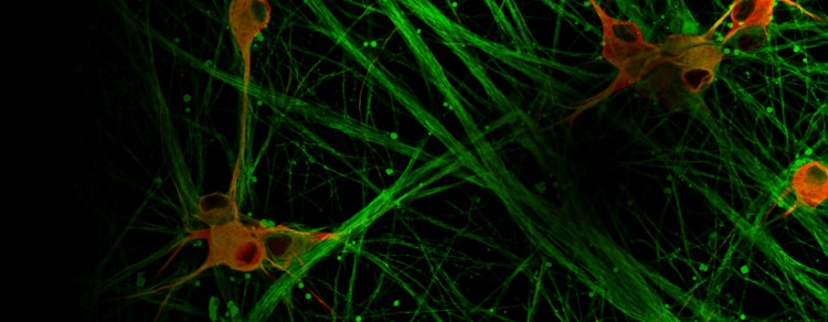
High-Content Imaging & Analysis
Accelerate discovery with a unified platform that combines automated imaging, advanced analysis, and AI-driven insights to streamline high-content screening workflows.
Uncover phenotypic insights at scale
High-content imaging and analysis combine automated microscopy with advanced, AI-driven data analysis to capture and extract phenotypic insights from complex cellular systems.
This integrated approach enables researchers to measure, quantify, and interpret multiple cellular features across large datasets—accelerating discovery from image acquisition to biological insight.
A Unified High-Content Screening Solution
Molecular Devices delivers an end-to-end platform that seamlessly integrates:
- High-resolution imaging
- AI-powered analysis
- Phenotypic interpretation
- Eliminating fragmented workflows, this unified solution enables researchers to move faster from data capture to actionable insight.

End-to-End Workflow
Capture, analyze, and interpret cellular data without switching platforms.

AI Powered Analysis
Automate segmentation, classification, and phenotypic profiling with advanced algorithms.

Performance that Keeps Pace
Scale from single-assay work to higher-volume screening with consistency you can rely on.

Reproducible Insights
Standardized workflows ensure reliable, publication-quality results.
How It Works: Capture Analyze Interpret
Scalable Imaging for High-Content Discovery
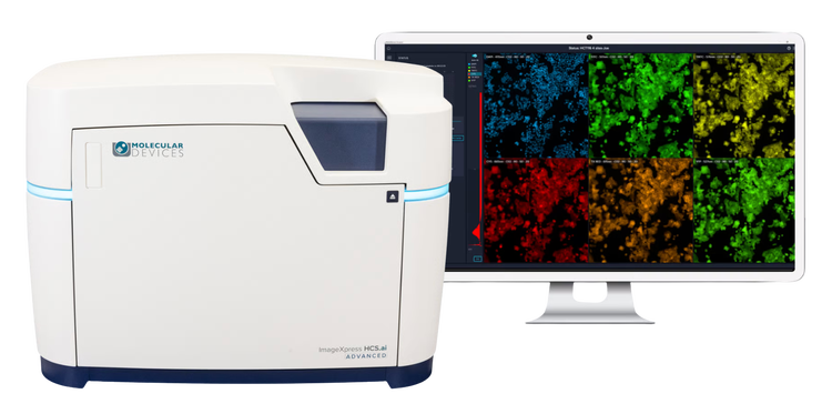
Our ImageXpress® systems support routine automated microscopy to advanced high-throughput confocal systems enabling scalable, powerful, Powered by AgileOptix™ technology, advanced illumination and optics, scientific sCMOS detection, and rapid acquisition, researchers can capture high-quality cellular data with speed, precision, and consistency.
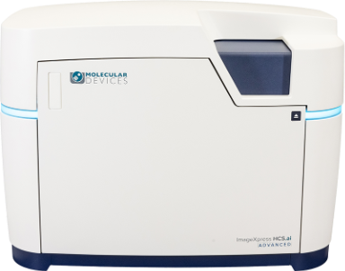
Capture: Acquire high-quality images with confidence
MetaXpress® Acquire software simplifies image acquisition with intuitive workflows, guided protocol setup, and flexible assay configuration. From routine screening to complex 3D and live-cell experiments, it helps users generate consistent, high-quality image data with the ImageXpress HCS.ai system.
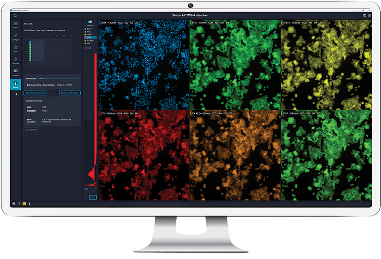
Analyze: Unlock deep insights from high-content imaging
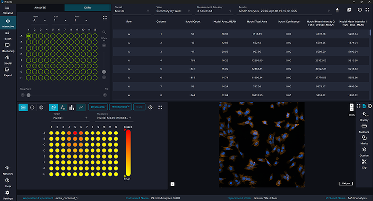
AI-powered analysis transforms complex images into precise, quantitative data, enabling faster, more confident decisions. Our integrated software combines advanced segmentation, feature extraction, and machine learning to deliver accurate, reproducible results at scale.
Measure and characterize:
- Cell morphology for phenotypic profiling
- Protein expression with quantitative precision
- Subcellular localization for deeper insight
- Cellular responses to perturbations across conditions
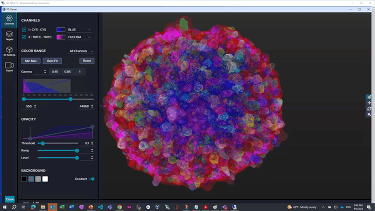
Interpret: From images to insight, at scale
Integrate imaging and analysis to generate rich, multiparametric phenotypic profiles across 2D and complex 3D models.
- Capture subtle phenotypic shifts others miss
- Decode complex biology with high-dimensional data
- Accelerate discovery from experiment to decision
Built for your research needs
Pharmaceutical Organizations
Drive high-throughput screening and large-scale discovery programs with integrated, enterprise-ready solutions.
Key Benefits:
- Process large volumes of samples and data at scale
- Improve reproducibility across teams and sites
- Accelerate compound screening and validation
Typical Use Cases:
- High-throughput screening (HTS)
- Toxicology studies
- Target identification and validation
Biotech Companies
Accelerate innovation with scalable, automated workflows that enable rapid hypothesis testing and data-driven decisions.
Key Benefits:
- Streamline assay development and optimization
- Scale screening workflows efficiently
- Leverage AI to extract deeper biological insights
Typical Use Cases:
- Phenotypic screening
- Functional genomics
- Early-stage drug discovery
Academic Researchers
Advance fundamental biology and translational research with flexible, high-resolution imaging and powerful analysis tools.
Key Benefits:
- Explore complex cellular mechanisms and pathways
- Perform detailed phenotypic characterization
- Support diverse assay types and experimental designs
Typical Use Cases:
- Cell biology research
- Disease modeling
- 3D biology and organoids
FAQs
What is high-content imaging and why is it important for drug discovery?
High-content imaging (HCI) uses automated microscopy and quantitative image analysis to measure multiple cellular features from the same experiment. Instead of producing a single readout, HCI turns cellular images into rich, multiparametric data that can help researchers understand how cells respond to compounds, genetic perturbations, disease models, or environmental conditions.
In drug discovery, this is valuable because researchers can evaluate biology in a more visual and information-rich way. HCI can support phenotypic screening, target validation, mechanism-of-action studies, toxicity assessment, and hit-to-lead decision-making by preserving cellular and subcellular context that single-endpoint assays may miss
Molecular Devices supports these workflows with ImageXpress® high-content imaging systems, MetaXpress® Acquire Software for image acquisition, and IN Carta® Image Analysis Software for AI-enabled image analysis, helping researchers move from image capture to quantitative, decision-ready results.
What is the difference between high-content imaging and high-content screening?
The terms are closely related, but they emphasize different parts of the workflow:
• High-content imaging (HCI) focuses on acquiring detailed cellular images and extracting quantitative information from them.
• High-content screening (HCS) applies HCI and HCA at screening scale, often across muliple plates, many compounds, genetic perturbations, doses, time points, or biological conditions and often includes increased throughput with automation.
In practical terms, HCI helps researchers see and quantify complex biology, while HCS uses that capability in a higher-throughput experimental workflow. Molecular Devices provides an integrated path from acquisition to analysis through ImageXpress® imaging platforms, MetaXpress® Acquire Software, and IN Carta® Image Analysis Software
How does AI improve high-content image analysis?
AI-enabled image analysis can help researchers handle the scale and complexity of high-content imaging data. Modern HCI and HCS experiments may generate large image datasets with many fields of view, channels, wells, plates, and biological conditions. AI and machine-learning tools can help segment cells or 3D structures, identify patterns, classify phenotypes, and extract quantitative measurements more efficiently than manual review.
With IN Carta® Image Analysis Software, researchers can use tools such as SINAP for deep-learning-based segmentation and Phenoglyphs™ for machine-learning-based phenotypic classification. These tools are designed to help users analyze complex 2D, 3D, and 4D image datasets, quantify phenotypic changes, and explore subtle biological patterns in a guided workflow.
AI-enabled analysis is most powerful when paired with high-quality images, appropriate controls, thoughtful assay design, and validated analysis settings. It should be viewed as a way to improve consistency, scalability, and biological insight, not as a substitute for good experimental design.
Can high-content imaging or screening be used with 3D cell models and organoids?
Yes. High-content imaging is widely used to study 3D cell models, including spheroids, organoids, tumoroids, and other human relevant models. These models can better represent aspects of tissue architecture, cell-cell interactions, gradients, and drug responses than many traditional 2D monolayer cultures, making them useful for disease modeling, toxicology, and drug discovery research.
3D models also create new imaging and analysis challenges. Thick samples can require z-stacks, confocal imaging, higher sensitivity, careful staining, and advanced segmentation methods to extract reliable quantitative data. Molecular Devices supports 3D biology workflows through ImageXpress® systems and IN Carta® analysis tools that can help researchers acquire, segment, quantify, and interpret complex 3D image datasets.
For best results, researchers should match the imaging modality, magnification, optical sectioning, live-cell requirements, and analysis workflow to the biology of the model and the decision the assay needs to support.
How do I choose the best high-content imaging system for my research?
The best high-content imaging system depends on your biology, assay format, throughput needs, imaging requirements, analysis workflow, and long-term growth plans. Key questions include:
• Are you working with 2D monolayers, 3D spheroids, organoids, tissues, or live-cell models?
• Do you need widefield imaging, confocal imaging, or both?
• How many plates, wells, fields, channels, or time points do you need to capture?
• What level of resolution and sensitivity performance does your assay require?
• Do you need automation, environmental control, or support for multi-user core facility workflows?
• How important are AI-enabled segmentation, phenotypic classification, batch analysis, and downstream data management?
• Do you need a modular system that can expand as applications become more complex?
Molecular Devices offers a range of ImageXpress® high-content imaging solutions, including systems designed for automated cell imaging, advanced confocal imaging, and high-throughput screening workflows. The ImageXpress® HCS.ai High-Content Screening System combines configurable imaging hardware with MetaXpress® Acquire Software and IN Carta® Image Analysis Software, giving laboratories a scalable acquisition-to-analysis workflow for both routine assays and more advanced 2D and 3D applications.
A practical next step is to start with the biological question and work backward: define the sample type, readouts, throughput, imaging modality, analysis needs, and decision point, then select the system configuration that best supports that complete workflow.
Latest Resources
Featured cell imaging applications
Cancer Research
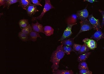
Cancer involves changes which enable cells to grow and divide without respect to normal limits, to...
Cell Imaging
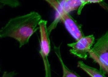
Researchers have several options in methods for imaging cells, from phase-contrast microscopy that...

3D Cell Models
Development of more complex, biologically relevant, and predictive cell-based assays for compound..
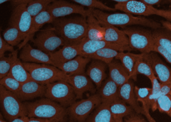
Live Cell Imaging
Live cell imaging is the study of cellular structure and function in living cells via microscopy....
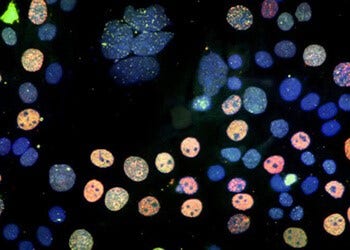
Cell Counting
Here we discuss the various methods and techniques used to assess proliferation, cytotoxicity, or...
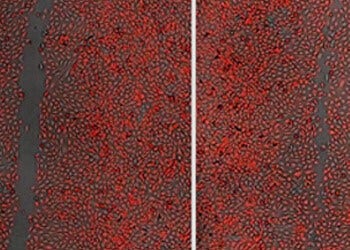
Cell Migration Assays
The movement or migration of cells is often measured in vitro to elucidate the mechanisms of...
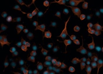
Neurite Outgrowth
Neurite outgrowth is assessed by the segmentation and quantification of neuronal processes. These...
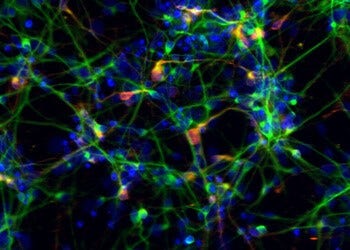
Stem Cell Research
Stem cells provide researchers with new opportunities to study targets and pathways that are more...