Cell Imaging & Analysis
Acquire and analyze quantitative data for cell-based model applications
Cell imaging: Acquisition and analysis
Researchers have several options in methods for imaging cells, from phase-contrast microscopy that shows intact cells to fluorescent imaging of single molecules or organelles. Cellular analysis is performed to evaluate and measure the current state of cells, such as cell integrity, toxicity, and viability and various other research applications. An integral part of cellular analysis is data collection, analysis, and export into a meaningful and useful format.
Cellular analysis workflow
Protocols for culturing, plating, and maintaining cells will vary between different cell models however, the steps below outline a general workflow for cell-based imaging assays from upstream media prep through downstream experimental analysis using a high-content imaging system and cellular analysis software.

- Plate cells into labware of choice (chamber slides, dishes, microplates)
- Treat cells – Perform cell treatments with desired compounds if part of the assay workflow
- Stain for markers – If required, label cells with the desired fluorophores (fluorescent dyes, fluorescent protein – peptide fusions, etc.
- Acquire cell image – Place the plate into the cell imaging system and start acquiring your live cell images
- Analyze cell image – Use cellular imaging analysis software to run quantitative analysis of live cell image
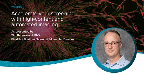
Accelerate your screening with high-content and automated imaging
This technical webinar will focus on high-content screening applications such as those involved in viral research, neurite outgrowth studies, and organoid models. The information provided will apply to researchers currently working with cell-based assays and doing live-cell imaging, and those looking to automate and/or increase their current imaging workflow.

Types of cellular imaging methods
Whether the demand is for a high-content and high-throughput or a simple-to-use system, our cellular imaging solutions can accommodate acquisition and analysis that suits the needs of a screening or research lab. Here, we’ve briefly described several methods in cellular imaging:
- Phase-contrast microscopy shows intact cells
- Fluorescent imaging can show single molecules or organelles
- Brightfield microscopy is useful for visualizing whole cells rapidly
- Confocal microscopy is preferred for high-quality images and 3D reconstruction of cells
- 4D live-cell imaging combines confocal with time-lapse microscopy
- Automated microscopy employs software and hardware features which ensure robust focus in every field-of-view combined with sophisticated image analysis software to generate repeatable and subjective quantitative data
Common cell-based assays and applications
Here, we’ve highlighted several cell-based assay topics from 3D cell cultures to stem cell research as well as applications for cardiomyocytes and toxicity screening.
Latest Resources
Cell Imaging & Analysis Workflow
Cell Imaging & Analysis Workflow

PLATE CELLS
Plate adherent or suspension cells into the labware of choice including culture dishes, chamber slides, and microplates. Incubate the cells at normal culture conditions (i.e. 37°C, 5% CO2). For adherent cells, incubating the plated cells overnight will allow ample time for cell attachment and growth.

96- or 384- Well Plates
Most cell cultures for high-content, high-throughput screening are run using 96- or 384-well microplates.

TREAT WITH COMPOUNDS
If necessary for the protocol, treat the cells with compounds of interest.
Compounds can be added to cells for minutes up to several days depending on the mechanism of action of the compounds and the biological re-sponse being investigated. For example, apoptosis or receptor internalization assays generally require shorter incubation periods while multiparametric cytotoxicity assays require longer periods. Longer compound treatments may require the replacement of compounds during incubation

STAIN FOR MARKERS
Live cells can be stained with a variety of fluorophores or transfected/transduced with constructs containing fluorescent protein – peptide fusions. Manufacturer instructions should be followed when staining. Stain-ing isn’t necessary for all live cell applications and label free imaging and analysis allows for the tracking and monitoring of cells in brightfield only.

Assay Kits
Easy-to-use, robust assay kits for life science research, drug discovery and development, and bioassays. Our assay kits are optimized for use on our instruments. Screen more compounds earlier in drug discovery and enable characterization of a full concentra-tion-response profile of test compounds.

ImageXpress High-content Imaging Portfolio
Our systems for high-content imaging and analysis provide flexible scalability making it easy to evolve your system alongside your research. They feature options and modules to address your specific research including objectives, filters, imaging modes, and envi-ronmental conditions.

ACQUIRE LIVE CELL IMAGE
For continuous time-lapse acquisitions, configure the total length of time for the time series and the imaging intervals, i.e. image every hour for 24 hours.
Discontinuous time-lapse acquisitions can be per-formed, where the plate is taken to and from the incubator and imaged at different time points.

ANALYZE CELL IMAGE
Analyze the images with cellular imaging analysis software to generate multiparametric readouts for the biological responses that are being studied. Kinetic data can be generated and reviewed for time-lapse acquisitions.
https://share.vidyard.com/watch/ABJi1T62w9u2H6GTC59Uxx
Cellular Imaging Acquisition and Analysis Software
MetaXpress® High-Content Image Acquisition and Analysis Software is a comprehensive solution for high-content analysis featuring a tightly orchestrated and integrated workflow. The portfolio of application modules supports a range of needs from ease-of-use through to proprietary assay design. The software includes powerful and elegant tools for 2D and 3D imaging such as time lapse analysis. With the assistance of our acquisition setup wizard, you’ll be generating data in minutes.
