Organoids

Biologic research and disease modeling recapitulate complexity of real tissues

What are organoids?

Organoids are three-dimensional (3D) multi-cellular, microtissues derived from stem cells that are designed to closely mimic the complex structure and functionality of human organs like the lung, liver or brain. Organoids are multi-cellular and demonstrate a high order of self-assembly to allow for an even better representation of complex in vivo cell responses and interactions, as compared to traditional 2D cell cultures.

There are three distinct definitions that differentiate an organoid:

  • It's a 3D biological microtissue containing several types of cells
  • Represents the complexity, organization and structure of a tissue
  • And, resembles at least some aspect of a tissue’s functionality
Airway Organoid

Organoids are becoming increasingly important in the fields of cancer research, neurobiology, stem cell research, and drug discovery, since they allow for the enhanced modeling of human tissues. Derived from stem cells, organoids can be differentiated into a wide range of tissue types including liver, lung, brain, kidney, stomach, and intestine. Because these 3D microtissues mimic in vivo organs, they can provide researchers with greater insight into the mechanisms of human development and disease. For example, researchers can grow organoids from genetically modified cells to understand how specific gene mutations are linked to certain genetic disorders. Organoids can also facilitate the study of infectious diseases and host-pathogen interactions. Finally, the ability to use patient-derived organoids for drug screens and toxicity evaluations is enabling researchers to make further advancements in personalized medicine.

General workflow for organoid culturing and screening

Due to the growing complexity of organoids and other 3D cellular systems, more sophisticated 3D imaging and analysis techniques are needed to accurately and efficiently characterize these biological assays. Today, automated confocal imaging systems and 3D image analysis software are commonly used to help researchers streamline their workflow and obtain optimal results.

Organoids workflow illustration

Step 1) 2D preculture

Organoids are derived from either primary cells (i.e., intestine, lung, or kidney) or induced pluripotent stem cells. Stem cells are able to differentiate and self-assemble into a variety of tissue-specific organoids.

Step 2) Developing 3D organoids

Typically cells are premixed with Matrigel and droplets are placed into a 24-well plate at room temperature. The plates are then placed into an incubator to form a solid droplet dome. Media is then added for seven or more days to promote cell growth and differentiation into specific tissue (brain, gut, lungs, etc.). Media includes ECM proteins, and different growth factors, which will vary depending on the type of tissue that is being developed.

Step 3) Organoid culture

Organoid culture is a long process and may include several steps with different media. During this process, cell health needs to be monitored (imaging), typically used for understanding developmental biology and tissues.

Step 4) Monitoring and readouts

Before experiments are conducted, organoids need to be monitored and characterized to ensure that they have the appropriate tissue structure and differentiation. High-content imaging allows for monitoring and visualizing the growth and differentiation of organoids, 3D reconstruction of the structures, complex analysis of organoids structure, cell morphology and viability, as well as the expression of different cell markers.

Step 5) Confocal imaging and 3D analysis

Confocal imaging and 3D analysis of organoids allow visualization and quantitation of the organoids and the cells that make up the organoid. Characterization of multiple quantitative descriptors of organoids could be used for studying disease phenotypes and compound effects.

Confocal imaging and 3D image analysis of organoids

Organoids are very useful for disease modeling and assessment of compound effects. Automated imaging and analysis of organoids are important for the  quantitative assessment of phenotypic changes in organoids, as well as for increasing throughput of experiments and screens.


Confocal imaging and 3D image analysis of organoids


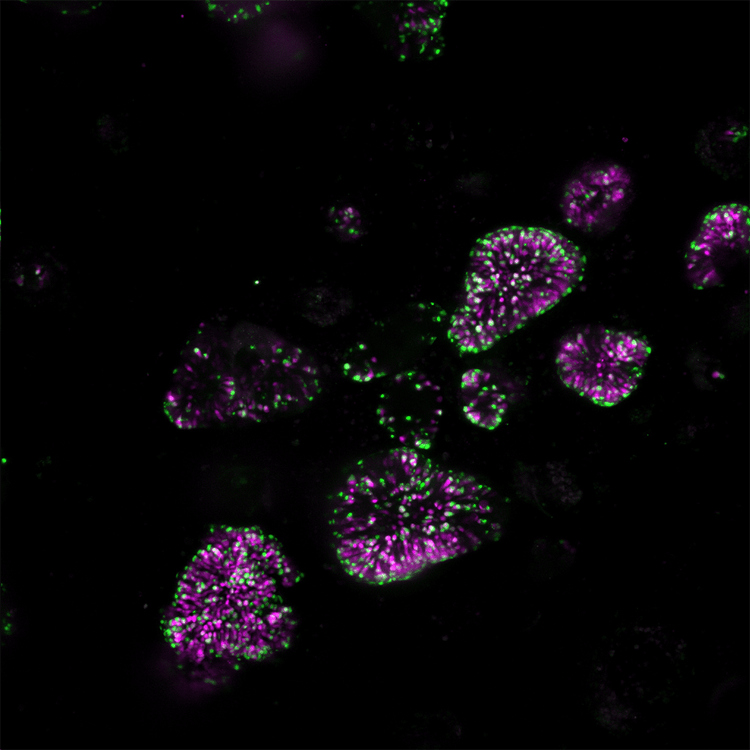
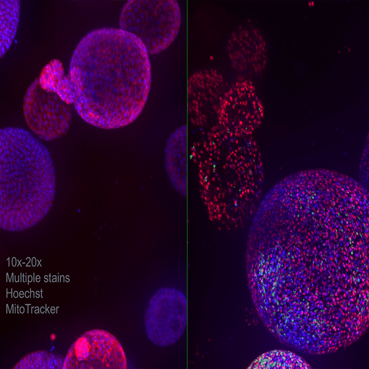
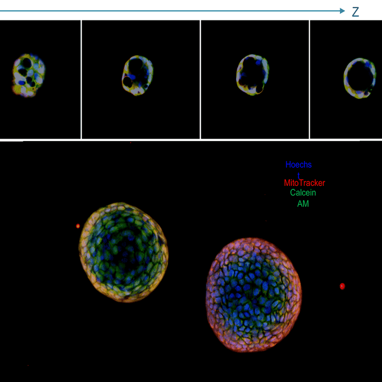
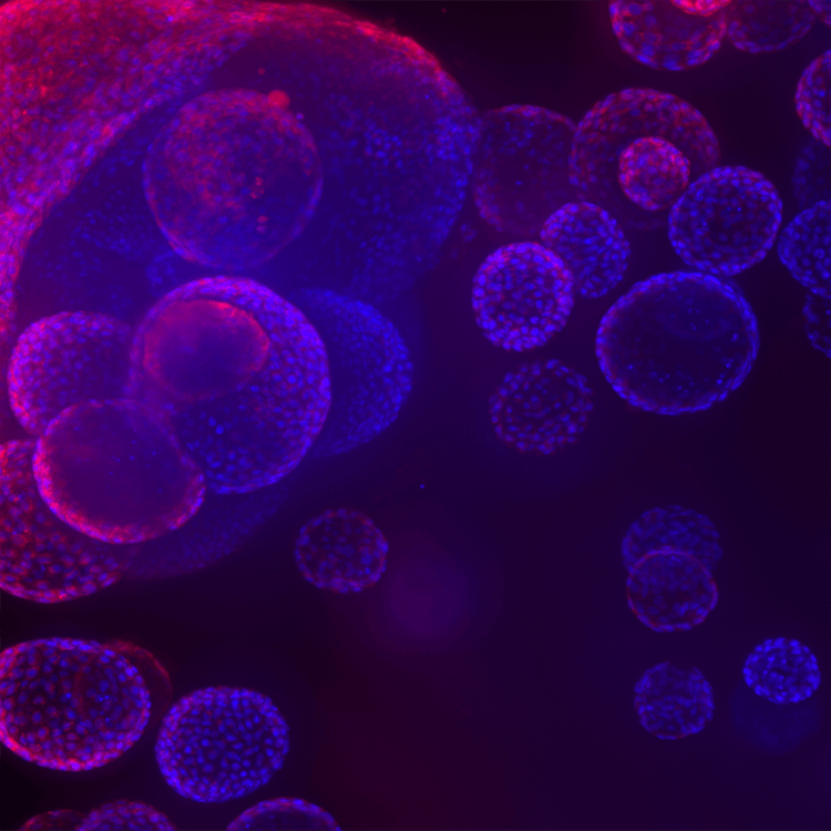
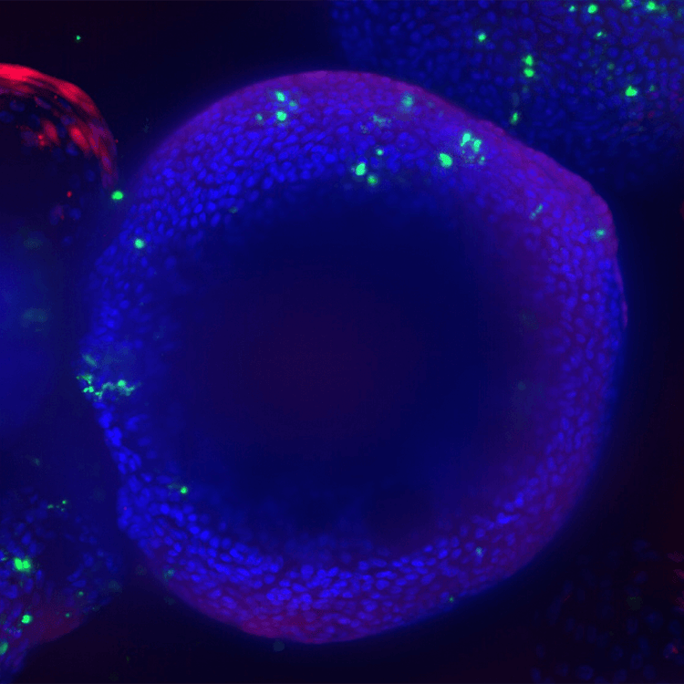
Confocal imaging, like the ImageXpress® Confocal HT.ai system with high-performance lasers and water immersion objectives are especially useful for capturing the complexity of 3D biological assays. Unlike typical spheroids, which have appearance of solid objects and limited penetration of light, some 3D organoids such as pulmonary organoids have a hollow appearance, with lumen, or cavities inside, and are more easily penetrated by light, allowing “imaging through” the microtissues embedded into Matrigel.

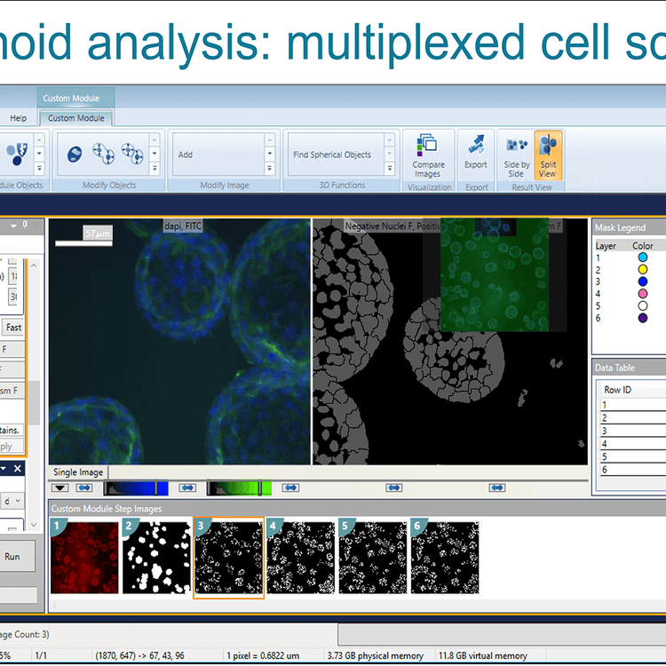
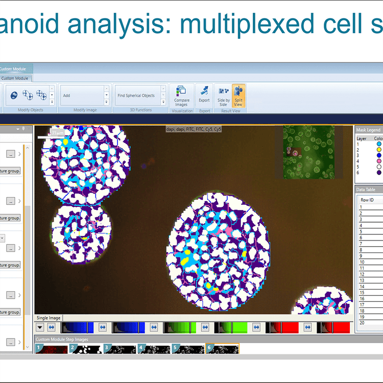
Airway Organoid CME analysis


Imaging and Analysis of a 3D Organoid Model. In this short video, Andy Bashford (Imaging Applications Scientist) shows a great example of a 3D Airway Organoid Model along with some interesting ways to get the most out of this type of assay.

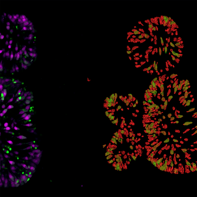
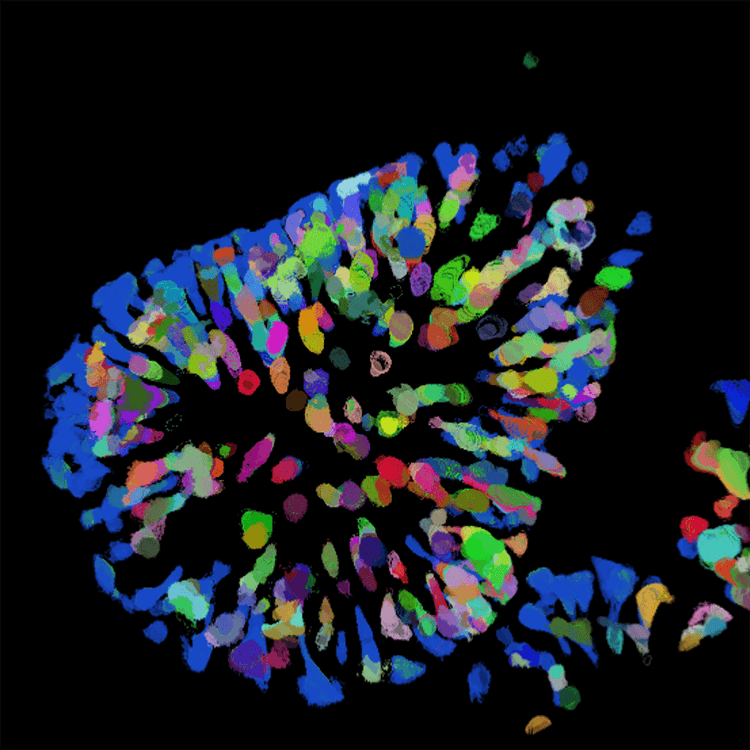
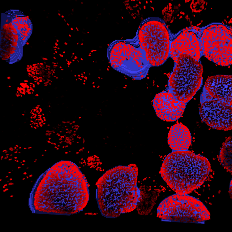
High-content analysis tools like the MetaXpress or IN Carta Image Analysis Software, tools allow for the finding and characterization of multiple objects/organoids, either in 2D format (for single plane, or maximum projection images) or in 3D when objects from the multiple planes are connected and reconstituted in 3D space by software. Organoids can be characterized for diameter, volume, shape, intensity of specific marker, or distance to other objects.

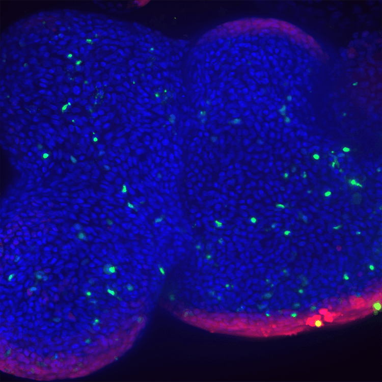
Furthermore, individual cells, nuclei, or organelles can be defined and measured within each organoid. That allows count of live and dead cells, or cells with a specific marker also defining volumes and distances between the objects. Numeric values can be counted for each organoid or averaged per well.

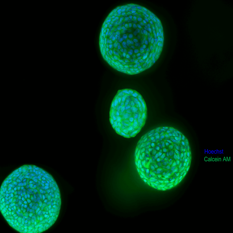
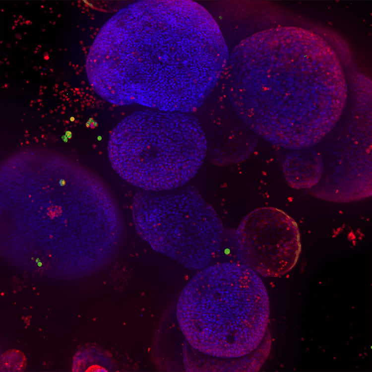
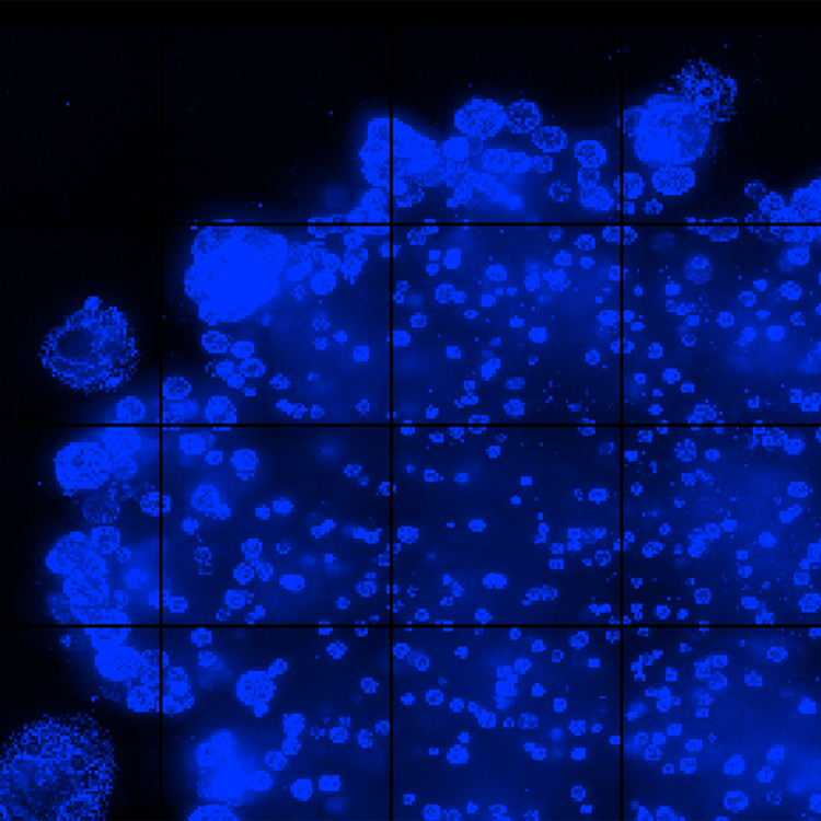
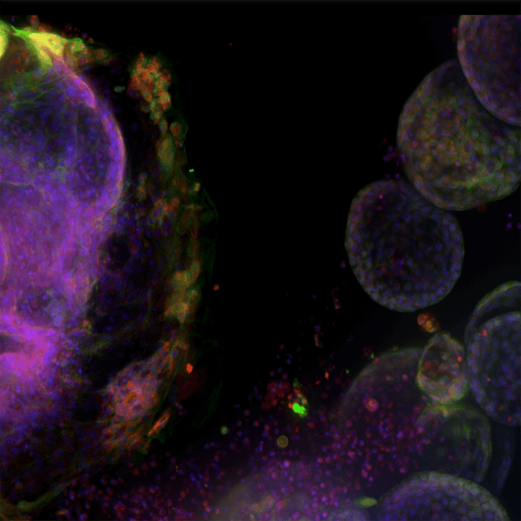
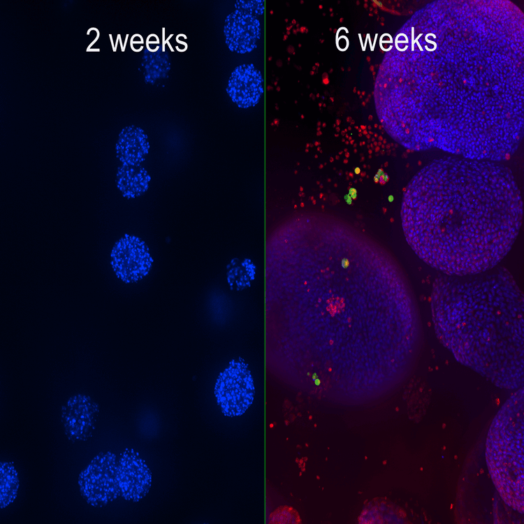
Lung organoid cell image gallery
Learn more about organoid research

  • 3D Cancer Cell Research

    3D Cancer Cell Research

    Cancer involves changes which enable cells to grow and divide without respect to normal limits, to invade and destroy adjacent tissues, and ultimately to metastasize to distant sites in the body. Cancer spheroids mimic tumor behavior far more effectively than standard 2D cell cultures. Such 3D spheroid models are being successfully used in screening environments for identifying potential cancer therapeutics.

    Cancer researchers need tools that enable them to more easily study the complex and often poorly understood interactions between cancerous cells and their environment, and to identify points of therapeutic intervention.

    Learn More 

    Brain Organoids

    Brain (Cerebral) Organoids

    Brain organoids are 3D tissue models representing one or more regions of the brain. They can overcome the shortcomings of conventional post-mortem and animal brain models to produce clinically relevant results.

    Cerebral organoids have great potential for understanding brain development and neuronal diseases. They can also be used for investigating genetic disorders and the effects of compounds. Nevertheless, capturing the uniqueness of the human brain requires functional assays and high-content imaging systems.

    Learn more about brain organoids 

  • Drug Discovery & Development

    Drug Discovery & Development

    The drug discovery landscape is shifting, with more scientists centering cell line development, disease models, and high-throughput screening methods around physiologically-relevant 3D cell models. The reason for this is clear: Using cellular model systems in research that closely mimic patient disease states or human organs can bring life-saving therapeutics to market – faster.

    Learn more  

    Intestinal Organoids

    Intestinal Organoids

    Intestinal organoids are 3D tissue models that recapitulate structures in the intestinal lumen and on the surrounding intestinal epithelium.

    The cell composition and arrangement of the epithelium make intestinal organoids useful for studying intestinal cell biology, regeneration, differentiation, as wells as diseases phenotypes including effects of specific mutations, microbiome, or inflammation process.

    Learn more about intestinal organoids 

  • Lab Automation for Organoid Screening Workflow

    Lab Automation for Organoid Screening Workflow

    Our lab automation solutions include scientists and engineers who can customize our instruments, as well as automate entire workflows to meet the specific needs of your assay, method, or protocol. From incubators, liquid handlers, and robotics to customized software and hardware—and with over 35 years of experience in the life science industry—you can count on us to deliver quality products and provide worldwide support.

    Learn more about how robotics-driven automation workcells and AI-based image analysis can help you develop an efficient, end-to-end workflow for your organoid development process.

    Patient-derived organoids (Tumoroids)

    Patient-derived organoids

    Patient-derived tumor organoids or tumoroids are cultures of tumor cells that can be generated from individual patients. Tumoroids are highly valuable tools for cancer research, drug development, and personalized medicine.

    Early detection and treatment are crucial in the survival rate of breast cancer patients. This necessitates the use of clinically relevant tumor models to understand the mechanism, analyze tumor biomarkers, and screen anticancer drugs. Breast cancer tumoroids provide the platform to study tumor physiology and response to targeted therapies.

    Learn how to analyze breast cancer tumoroid growth and the efficacy of anticancer treatments with high-throughput screening and high-content imaging solutions:

    Learn more about patient-derived breast cancer tumoroids 

  • Pulmonary (Lung) Organoids

    Pulmonary (Lung) Organoids

    Lung organoid cultures are 3D microtissue models recapitulating the morphological and functional characteristics of the airway, such as mucus secretion, ciliary beating, and regeneration. This biological relevance enables the study of repair/regeneration mechanisms in lung injury and phenotypic changes in pulmonary diseases. Lung organoids also can be used for toxicity assessment or drug testing.

    Learn more about lung organoids 

    Spheroids

    Spheroids

    Spheroids are multi-cellular 3D structures that mimic in vivo cell responses and interactions. They can be highly reproducible and to be scaled for high-content screening. Compared with adherent cells grown in 2D monolayers, 3D growth conditions are believed to more closely reflect the natural environment of cancer cells. Acquiring measurements from these larger structures involve acquiring images from different depths (z-planes) within the body of the spheroid and analyzing them in 3D, or collapsing the images into a single 2D stack before analysis.

    Learn More 

  • Stem Cell Research

    Stem Cell Research

    Pluripotent stem cells can be used for studies in developmental biology or differentiated as a source for organ-specific cells and used for live or fixed cell-based assays on slides or in multi-well plates. The ImageXpress system has utility in all parts of the stem cell researcher’s workflow, from tracking differentiation, to quality control, to measuring functionality of specific cell types.

    Learn more 

    Toxicology

    Toxicology

    Toxicology is the study of adverse effects of natural or man-made chemicals on living organism. It is a growing concern in our world today as we are exposed to more and more chemicals, both in our environment and in the products we use.

    Read More 

Latest Resources

Resources for Organoids

Videos & Webinars

Scale Up Your Screening: Integrating Automation Into 3D Biology for Therapeutic Drug Discovery

Are Organoids the Future of Drug Discovery?

Using Differentiated iPSCs to Build Ready-to-use 3D Models

The search for answers: Using lab automation with patient-derived tumoroids to find more relevant therapies for clinically aggressive cancers

New standard in organoid culture and image-based analyses

The BAB400 integrated with the ImageXpress® Confocal HT.ai High-Content Imaging System

The BAB400 integrated with the ImageXpress® Confocal HT.ai High-Content Imaging System

Leverage automated, end-to-end workflows to enable complex organoid assays

Enhance high-content 3D biology imaging with automated sample preparation

Enhancing 3D Disease Models: Automated, High-Throughput, Phenotypic Screening with Organ-on-a-Chip

Emerging Organoid Models: Translating Basic Research to Drug Development and Regenerative Medicine

Application and Analysis of Organoid Systems

Automating culture and high-content imaging of 3D organoids for in vitro assessment of compound effects

Getting Started with 3D Human Tissue Models and Imaging

Capturing the Complexity of Cell Biology

Monitoring organoid development in iPSC-derived 3D cerebral organoids

ISSCR 2021 Innovation Showcase: Automated Culture and High-Content Imaging of 3D Lung and Cardiac

Organoid Innovation Center Walkthrough

Organoid Innovation Center Walkthrough

Capturing the complexity of 3D biology: Organoids for disease modelling and toxicity research

Disease modeling in the 21st century: Automated organoid assays with 3D imaging

High-throughput, organoid-derived organ-on-a-chip systems for drug discovery and disease modelling

High-throughput, organoid-derived organ-on-a-chip systems for drug discovery and disease modelling

Developing high-throughput organ-on-a-chip tissue models for drug discovery using high-content imaging

Physiologically-Relevant Tissue Models Using a High-Throughput Organ-on-a-Chip Platform

Physiologically-Relevant Tissue Models Using a High-Throughput Organ-on-a-Chip Platform