
Get to Know our Field Applications Scientist: Cheryl Bell
Cheryl Bell talks about making the transition from manual microscopy to automated microscopy

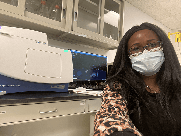
Cheryl, please tell us about your background and how you arrived at Molecular Devices.
I completed my undergraduate studies at Jackson State University in Mississippi, where I majored in pre-med biology. I took a genetics class as part of the curriculum and absolutely loved it! This led me to pursue my doctorate degree in Genetics and Genomics at the University of Connecticut where my focus was on HIV prevention therapy for uncircumcised males. The project was linked to a clinical trial that required years of microscopic evaluation of the samples to validate the efficacy of the therapeutic drug that we were studying. From there, I completed my postdoc training in cell biology at the University of Pittsburgh where I studied gap junction proteins with the intent to apply my learnings to the field of wound healing.
Throughout the course of my studies, I employed different types of microscopy techniques including fluorescence, confocal, and transmission electron microscopy. My goal at that time was to become a professor and run my own lab. Toward the end of my postdoc training, my plans took a turn when one of my friends from graduate school contacted me about an open position for a Cellular Imaging Field Applications Scientist at Molecular Devices. I ended up getting the position in April 2018.
Making the transition from academia to a corporate setting allowed me to transfer the knowledge and skill sets that I picked up from my years of research into helping customers answer their scientific questions through microscopy.
Can you tell us more about your role at Molecular Devices?
I am currently based out of Chicago, Illinois. In my role as a Field Applications Scientist, I provide scientific and sales support for our ImageXpress® Pico Automated Cell Imaging System. This includes conducting educational seminars or one-on-one sessions with customers, where I answer technical questions related to the product or specific applications.
The ImageXpress Pico system was launched around the same time that I joined Molecular Devices. Since then the platform has evolved quite a bit, with enhancements such as Environmental Control, Digital Confocal, and Live Preview.
Are most ImageXpress Pico system customers novice or advanced microscopy users?
I would say that half of our customers are novice users who were accustomed to using manual microscopy prior to transitioning to the ImageXpress Pico system. These customers were looking to increase throughput and decrease the amount of time that’s spent acquiring images and analyzing data. Using this system, these customers are now able to cut the imaging portion of their workload from hours to minutes. They are also able to generate more data from their experiment.
Typical functions that you would have to perform on a manual microscope are streamlined with automated imaging. This eliminates the need to make adjustments during image acquisition. Essentially, the ImageXpress Pico system helps customers optimize their cellular imaging and analysis workflow.
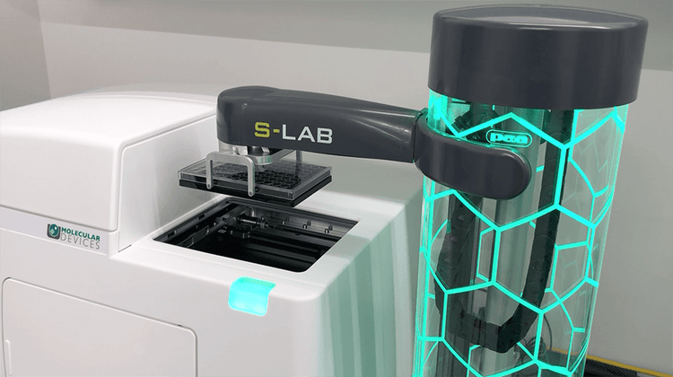
I would consider the remaining portion of our customers to be advanced users who are already familiar with automated imaging. These individuals look to the ImageXpress Pico system for its ease-of-use, high-resolution image quality, as well as the versatility that it offers in accommodating different sample types and labware. More advanced users usually take advantage of our Customization and Automation solutions. This may involve upgrading to additional features in the CellReporterXpress® software or combining with other Molecular Devices software such as MetaXpress® High-Content Image Acquisition and Analysis Software or SoftMax® Pro Software. Within these software platforms, they can activate certain functions such as Z-stack acquisition or Digital Confocal. In addition, they can use our newest customizations with a robotic arm and incubator.
In essence, the ImageXpress Pico system is designed to accommodate a wide range of users. Because the instrument is so easy to use, it is ideal for facilities with high turnover -- allowing new staff to get up and running very quickly.
What are some of the common challenges that you observe with customers as it relates to automated imaging?
I think that a common challenge for our customers is managing data. Automated imaging generates a lot of data. This can be on the scale of gigabytes or even terabytes. In many instances, we have to guide customers on how to save the data into different formats that are compatible with their systems.
Another challenge for customers is determining the types of plates or stains that are optimal for their particular application or assay. For instance, the way customers are accustomed to preparing their samples may not be ideal for automated imaging. I work with customers to make the necessary adjustments in sample preparation so that they can easily automate the imaging process and generate the data they need.
I’ve also observed that customers are becoming more interested in imaging 3D cell models. In these cases, we help guide them to identify the imaging system that would best meet their research needs. Our imaging portfolio is configured in a way that makes it easy for customers to transition from the ImageXpress Pico system to a higher model like our latest ImageXpress® Confocal HT.ai High-Content Imaging System as their research continues to evolve.
Are there instances in which customers may choose to use the ImageXpress Pico system alongside the ImageXpress Confocal HT.ai system as part of their workflow?
Yes, definitely. We have customers who use the ImageXpress Pico system to perform validation testing or more straightforward imaging, where they need to run 96-well plates at high throughput. When customers need to acquire higher resolution images for a specific application, they can run the plate in the ImageXpress Confocal HT.ai system. The ease of using the ImageXpress Pico system helps clients to get through their experiments more quickly and ultimately acquire the data that they need.
We also have customers who use our microplate readers alongside the ImageXpress Pico system for certain applications. You can easily transfer the data acquired from the ImageXpress Pico system to SoftMax Pro Software which is used with our microplate readers. I think of the ImageXpress Pico system as the intermediary platform that helps our clients get to where they need to go in a more efficient manner.
Are there any particular applications or assays that really excite you as it pertains to imaging?
What I love most about my role as an applications scientist is that I get to see our systems being used for a wide range of applications. The ability to image organisms such as worms, phytoplankton, rotifers, and see molecular biology at its core in real time is very exciting me! I also get excited about working with samples such as mouse brain and zebrafish.
I’m seeing customers develop some groundbreaking assays through their customization of labware. They can create all types of labware designed to grow certain types of cells or allow bacteria to grow communities. As an example, I have customers who are developing multi-channels for neurons to grow. This creates an environment which is more representative of how neurons operate in vivo. Because these platforms are more biologically relevant, they enable researchers to probe further into their samples and design better drugs for different conditions such as Alzheimer’s. I have another customer who is studying bacterial communities in the gut so that they can gain greater insight into how to design probiotics for individuals who are suffering from gastrointestinal issues. The innovative ways in which customers are creating real biological scenarios to improve the way we understand health and disease truly blows my mind!
Are your imaging systems compatible with the customized labware that customers are developing?
Our imaging systems are compatible with a wide variety of labware. When needed, our team will work with customers to configure our hardware and/or software platforms to accommodate their unique labware requirements.
https://share.vidyard.com/watch/261PZTTCKAUFtD7skdrw2M
What future advancements do you envision for automated microscopy?
Automated microscopy will open a lot of avenues for different areas of research. For example, if you are in the organoid field, you are growing these samples over time. There is the need to repeatedly image the samples at different times during the growth process. I recently imaged a sample that was 160 days old. As we image samples that are increasing in size, we will need platforms that can see through these types of structures. I foresee that we will need to develop technology that is an intermediary between microscopy and CAT scanning. This would allow us to look even deeper into the sample.
Along these lines, I think the ImageXpress Pico system will continue to evolve and we will see enhancements that help further improve assay development and streamline the imaging workflow.
Looking back at your career, was there something in particular that sparked your interest in science?
From childhood I was interested in the inner workings of the human body particularly as it relates to normal and diseased states. I think this curiosity stemmed from my sister, who developed an autoimmune condition as a teenager. I would ponder what could cause an individual such as my sister to be healthy one day and then sick the next day. This really piqued my interest in science.
What are some of your interests outside of work?
I enjoy spending time with my three children. Now that they are older, I find that I have more time to pick up new hobbies. I would consider myself an ambivert. When I’m out with friends or working with colleagues, I’m very outgoing. When I’m at home, I like to spend some alone time reading or listening to music. One of my favorite everyday activities is running. I find it to be a form of meditation for me and I’m proud to have completed my first marathon last year.

To learn more about our automated imaging platforms, explore our Cellular Imaging Systems page.