High-content confocal imaging solution with water objective options


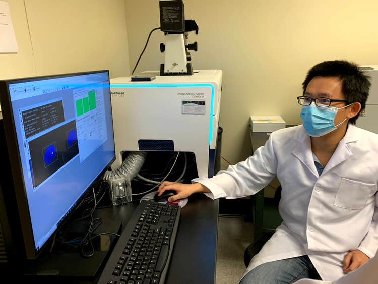
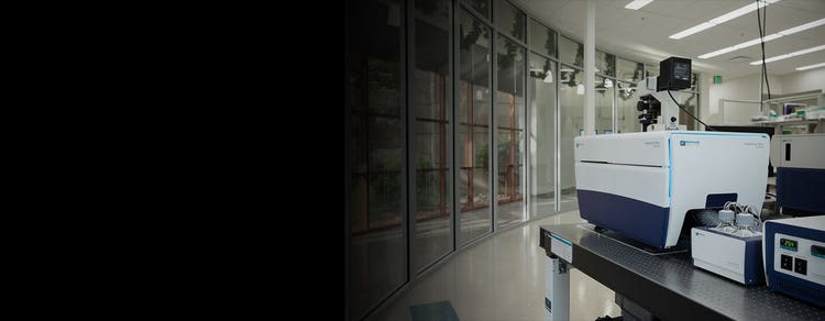
The ImageXpress® Micro Confocal system is a high-content solution that can switch between widefield and confocal imaging of fixed and live cells. It can capture high quality images of whole organisms, thick tissues, 2D and 3D models, and cellular or intracellular events. The spinning disk confocal and sCMOS camera enable imaging of fast and rare events like cardiac cell beating and stem cell differentiation. With MetaXpress software and flexible options like water immersion objectives to choose from, the system enables many confocal imaging applications from 3D assay development to screening.

  • Images Icon

    Acquire higher quality images

    Capture excellent contrast, high resolution images with our proprietary AgileOptix™ spinning disk confocal technology, wide field of view, and bright light source.

  • Customizable Icon

    Customize image acquisition and analysis

    Take ultimate control over acquisition and analysis parameters, which enables many applications from 3D structure analysis to target imaging of specific objects within an organism or cell populations. Run more applications with standard options like water immersion objectives, environmental control, or any of our high-performance customizations.

  • Analysis Icon

    Analyze more data in less time

    MetaXpress® PowerCore™ software accelerates analysis speed in a high-throughput environment. The software distributes image processing jobs to a multi-CPU environment.

ImageXpress Micro Confocal Virtual Tour

ImageXpress Micro Confocal Virtual Tour

Features

  • Breadth Icon

    Wide dynamic range

    Quantify low and high intensity signals in a single image with >3 log dynamic range intensity detection.

  • Optics Icon

    Exclusive AgileOptix spinning disk confocal

    The technology provides increased sensitivity with specially designed optics, high-powered, solid state light engines, and sCMOS sensor. Swappable disc geometries provide flexibility between speed and resolution.

  • Accuracy Icon

    Wide field of view

    Wide field of view enables whole-well confocal imaging and eliminates missed targets.

  • Automation Icon

    Optional on-board robotic fluidics

    For assays that involve compound addition, well washing, and media exchange, optional on-board robotic fluidics are available.

  • Measure Icon

    Accurate 3D measurements

    MetaXpress 3D analysis module is optimized for confocal imaging, enabling 3D measurements of volume and distance.

  • Expandable Icon

    Multiple imaging modes

    The system offers phase contrast and brightfield label-free imaging, fluorescence, widefield, colorimetric, and confocal imaging, as well as, water immersion imaging as a standard option.

AgileOptix™ technology

Our proprietary AgileOptix technology enables the ImageXpress Micro Confocal system to deliver the sensitivity and throughput needed for demanding applications. AgileOptix is the combination of a powerful solid state, light engine, specially designed optics, scientific CMOS sensor, and the ability to change disk geometries.

AgileOptix Technology for ImageXpress Micro Confocal


Introducing the MetaXpress 3D Analysis Toolkit

MetaXpress high-content image aquisition and analysis software


  • Meet high throughput requirements with a scalable, streamlined workflow
  • Adapt your analysis tools to tackle your toughest problems, including 3D analysis
  • Schedule automatic data transfer between third-party hardware sources and secure database
  • Set up hundreds of routinely used HCS assays using MetaXpress software modules


Cellular Image Gallery

Micro Confocal High-Content Imaging System
ImageXpress High-content imaging solution
ImageXpress Micro Confocal Imaging System
ImageXpress Micro Imaging System
Micro Confocal Imaging System
 
Data and images were acquired during development using customer samples. Results may vary. Highlighted features’ price, time to deliver, and specifications will vary based on mutually agreed technical requirements. Solution requirements may cause adjustment to standard performance.


Latest Resources

209

Applications of ImageXpress Micro Confocal High-Content Imaging System

  • 3D Cell Imaging and Analysis

    3D Cell Imaging and Analysis

    Three-dimensional (3D) cell models are physiologically relevant and more closely represent tissue microenvironments, cell-to-cell interactions, and biological processes that occur in vivo. Now you can generate more predictive data by incorporating technologies like the ImageXpress system with the integrated 3D Analysis Module in MetaXpress® software. This single interface will enable you to meet 3D acquisition and analysis challenges without compromise to throughput or data quality, giving you confidence in your discoveries.

    Learn More 

    Cancer Research

    Cancer Research

    Cancer researchers need tools that enable them to more easily study the complex and often poorly understood interactions between cancerous cells and their environment, and to identify points of therapeutic intervention. Learn about instrumentation and software that facilitate cancer research using, in many cases, biologically relevant 3D cellular models like spheroids, organoids, and organ-on-a-chip systems that simulate the in vivo environment of a tumor or organ.

    Learn More  

  • Cell Counting

    Cell Counting

    Cell counting is fundamental and critical to numerous biological experiments. Assays such as drug compound toxicity, cell proliferation, and inhibition of cell division rely on the assessment of the number or density of cells in a well.

    Learn More 

    Cell Migration Assays

    Cell migration assays

    The movement or migration of cells is often measured in vitro to elucidate the mechanisms of various physiological activities such as wound healing or cancer cell metastasis. Cell migration assays may be conducted in a controlled environment using live cell timelapse imaging.

    Learn More 

  • Cells in Extracellular Matrices

    3d-hydrogels

    One common way of culturing cells in three dimensional space is through the use of extracellular matrix-based hydrogels, such as Matrigel. Cells are grown in an extracellular matrix (ECM) to mimic an in vivo environment. Differences between Matrigel and 2D cell cultures can be readily seen by their different cell morphologies, cell polarity, and/or gene expression. Hydrogels can also enable studies on cell migration and 3D structure formation, such as endothelial cell tube formation in angiogenesis studies.

    COVID-19 and Infectious Disease Research

    COVID-19 and Infectious Disease Research

    Here we've addressed common applications in infectious disease research including cell line development, binding affinity, viral neutralization, viral titer and more with a focused effort on understanding the SARS-CoV-2 virus in order to develop potential therapies for COVID-19 including vaccines, therapeutics and diagnostics.

    Learn More 

  • Drug Discovery & Development

    Drug Discovery & Development

    The drug discovery landscape is shifting, with more scientists centering cell line development, disease models, and high-throughput screening methods around physiologically-relevant 3D cell models. The reason for this is clear: Using cellular model systems in research that closely mimic patient disease states or human organs can bring life-saving therapeutics to market – faster.

    Learn more  

    Live Cell Imaging

    Live Cell Imaging

    Live cell imaging is the study of cellular structure and function in living cells via microscopy. It enables the visualization and quantitation of dynamic cellular processes in real time. Live cell imaging encompasses a broad range of topics and biological applications—whether it is performing long-term kinetic assays or fluorescently labeling live cells.

    Learn More  

  • Neurite Outgrowth / Neurite Tracing

    Neurite Outgrowth

    Neurons create connections via extensions of their cellular body called processes. This biological phenomenon is referred to as neurite outgrowth. Understanding the signaling mechanisms driving neurite outgrowth provides valuable insight into neurotoxic responses, compound screening, and for interpreting factors influencing neural regeneration. Using the ImageXpress Micro system in combination with MetaXpress Image Analysis Software automated neurite outgrowth imaging and analysis is possible for slide or microplate-based cellular assays.

    Learn More  

    Organoid technology / Organ-on-a-Chip

    Organ on a Chip Assays

    Organoid technology, like organ-on-a-chip emulates organ physiology though co-culture of cells in a supportive 3D matrix and use of microfluidic channels to perfuse nutrients or compounds over the resulting cellular structures. It is rapidly gaining popularity as a biologically relevant screening model for new drugs and toxicity.

  • Spheroids

    Spheroids

    Spheroids are multi-cellular 3D structures that mimic in vivo cell responses and interactions. They can be highly reproducible and to be scaled for high-content screening. Compared with adherent cells grown in 2D monolayers, 3D growth conditions are believed to more closely reflect the natural environment of cancer cells. Acquiring measurements from these larger structures involve acquiring images from different depths (z-planes) within the body of the spheroid and analyzing them in 3D, or collapsing the images into a single 2D stack before analysis.

    Learn More 

    Stem Cell Research

    Stem Cell Research

    Pluripotent stem cells can be used for studies in developmental biology or differentiated as a source for organ-specific cells and used for live or fixed cell-based assays on slides or in multi-well plates. The ImageXpress system has utility in all parts of the stem cell researcher’s workflow, from tracking differentiation, to quality control, to measuring functionality of specific cell types.

    Learn more 

  • Toxicology

    Toxicology

    Toxicology is the study of adverse effects of natural or man-made chemicals on living organism. It is a growing concern in our world today as we are exposed to more and more chemicals, both in our environment and in the products we use.

    Read More 

Specifications & Options of ImageXpress Micro Confocal High-Content Imaging System

Resources of ImageXpress Micro Confocal High-Content Imaging System

Presentations
Videos & Webinars
Scale Up Your Screening: 3D Biology

Scale Up Your Screening: Integrating Automation Into 3D Biology for Therapeutic Drug Discovery

FUJIFILM Cellular Dynamics - Using Differentiated iPSCs to Build Ready-to-use 3D Models

Using Differentiated iPSCs to Build Ready-to-use 3D Models

Automated Imaging of 3D Assay Models

Explore More Complex Biology with Automated Imaging of 3D Assay Models

Immunology and Vaccine Development Workflow

Immunology and Vaccine Development Workflow

Hybridoma Workflow

Hybridoma Workflow

Disease modeling in the 21st century

Disease modeling in the 21st century: Automated organoid assays with 3D imaging

High-throughput, organoid-derived organ-on-a-chip systems for drug discovery and disease modelling

High-throughput, organoid-derived organ-on-a-chip systems for drug discovery and disease modelling

Unleash the power of Cell Painting

Unleash the power of Cell Painting

3D cell culture, tissue clearing, & high-content imaging

3D cell culture, tissue clearing, & high-content imaging in the quest of effective solutions to NAFLD

Transitioning high-content assays to 3D

Transitioning high-content assays to 3D: Scientific opportunities and imaging challenges

Water Immersion Objectives & High-Content Imaging

Gain deeper insights into cellular 3D structures with water immersion objectives

An AI-based approach to high-content phenotypic characterization of human iPSC-derived neuronal cells

An AI-based approach to high-content phenotypic characterization of human iPSC-derived neuronal cells

Implementing 3D Neural Spheroids

Implementing 3D Neural Spheroids in Drug Discovery

Accelerate your Screening with Automated Imaging

Accelerate your screening with high-content and automated imaging

Microplate based Detection

Accelerating study of viral infection and therapeutics with microplate-based detection and high-throughput screening

ImageXpress Micro Confocal Virtual Tour

ImageXpress Micro Confocal Virtual Tour

Magnetic 3D

Magnetic 3D Bioprinting, 3D Cell Culture in a 2D Workflow

LabTube Meets Molecular Devices & MIMETAS, Susan Murphy & Sebastiaan Trietsch

LabTube Meets Molecular Devices & MIMETAS, Susan Murphy & Sebastiaan Trietsch

Plate annotation and curve fitting in MetaXpress

Plate annotation and curve fitting in MetaXpress

Quickstart Guide: Plate acquisition on the ImageXpress Micro Confocal using MetaXpress

Quickstart Guide: Plate acquisition on the ImageXpress Micro Confocal using MetaXpress

Quickstart Guide: Review images on the ImageXpress Micro Confocal using MetaXpress

Quickstart Guide: Review images on the ImageXpress Micro Confocal using MetaXpress

Basic workflow from image acquisition to analysis on the ImageXpress Micro Confocal using MetaXpress

Basic workflow from image acquisition to analysis on the ImageXpress Micro Confocal using MetaXpress

Developing High Throughput Organ on a Chip Tissue

Developing high-throughput organ-on-a-chip tissue models for drug discovery using high-content imaging

Toxicity studies of iPSC-derived cardiomyocytes and neuronal spheroids

Toxicity studies of iPSC-derived cardiomyocytes and neuronal spheroids

3D in Vitro Models

Optimizing high-content screening tools for physiologically-relevant 3D in vitro models

Morphological Characterization of 3D Neuronal Networks in a Microfluidic Platform

Morphological Characterization of 3D Neuronal Networks in a Microfluidic Platform

3D Imaging of Cancer Spheroids

3D Imaging of Cancer Spheroids

High-Content Screening for Identifying miRNAs

High-Content Screening for Identifying miRNAs

Identification of Selective Inhibitors of the STAT3 Signaling Pathway

Identification of Selective Inhibitors of the STAT3 Signaling Pathway

Oliver Kepp and Jayne Hesley - Hallmarks of Cancer - Detect and Quantify Cell Death Signatures with High Content Imaging

Oliver Kepp and Jayne Hesley - Hallmarks of Cancer - Detect and Quantify Cell Death Signatures with High Content Imaging

Multidimensional High-Throughput Imaging with the New ImageXpress Micro Confocal System

Multidimensional High-Throughput Imaging with the New ImageXpress Micro Confocal System

Pushing the Boundaries of High Content Screening

Pushing the Boundaries of High Content Screening

Preparing Assays for Genome-wide RNAi Screens Using High Content Microscopy

Preparing Assays for Genome-wide RNAi Screens Using High Content Microscopy

Multiplexed High Content Hepatoxicity Assays Using iPSC-Derived Hepatocytes

Multiplexed High Content Hepatoxicity Assays Using iPSC-Derived Hepatocytes

High Content Imaging Analysis of Cell Sheet Morphogenesis Utilizing  𝘪𝘯 𝘷𝘪𝘵𝘳𝘰 Tissue Models

High Content Imaging Analysis of Cell Sheet Morphogenesis Utilizing 𝘪𝘯 𝘷𝘪𝘵𝘳𝘰 Tissue Models

Simple and Flexible High Content Imaging Enabling Quantitation of Complex Biological Events

Simple and Flexible High Content Imaging Enabling Quantitation of Complex Biological Events

Contemporary Automation and High Content Imaging Tools for Screening Stem Cell-Derived Cardiomyocytes

Contemporary Automation and High Content Imaging Tools for Screening Stem Cell-Derived Cardiomyocytes

Implement High-Throughput 3D Image Analysis for Samples from Subcellular Structures to Spheroids

Implement High-Throughput 3D Image Analysis for Samples from Subcellular Structures to Spheroids

Live Cell Imaging to Investigate the Regulation of Cell Division Timing

Live Cell Imaging to Investigate the Regulation of Cell Division Timing

Using High-throughput RNA Screening to Identify Host Factors That Impact Viral Infection

Using High-throughput RNA Screening to Identify Host Factors That Impact Viral Infection

Application of HCA Tools for Antibody Drug Discovery

Application of HCA Tools for Antibody Drug Discovery

Setting up 3D Spheroid Assays Using High-Content Imaging

Setting up 3D Spheroid Assays Using High-Content Imaging

Physiologically-Relevant Tissue Models Using a High-Throughput Organ-on-a-Chip Platform

Physiologically-Relevant Tissue Models Using a High-Throughput Organ-on-a-Chip Platform

Emerging Induced Pluripotent Stem Cell Applications in Drug Discovery

Emerging Induced Pluripotent Stem Cell Applications in Drug Discovery

StemoniX microBrain 3D Assay Ready Plates for HTS

StemoniX microBrain 3D Assay Ready Plates for HTS

  • Citation
    Dated: Jun 15, 2022
    Publication Name: American Association for Cancer Research (AACR) Journals

    Abstract 192: Ex vivo screening and analysis of novel effective treatments for ovarian cancer

    Purpose: Ovarian cancer is the most lethal gynecological cancer. Histologically, about 90% of the ovarian cancers have epithelial origin and more than 75% are characterized as high-grade serous ovarian cancer (HGSOC). View more

    Purpose: Ovarian cancer is the most lethal gynecological cancer. Histologically, about 90% of the ovarian cancers have epithelial origin and more than 75% are characterized as high-grade serous ovarian cancer (HGSOC).

    Contributors: Aikaterini Skorda; Kaisa Huhtinen; Wojciech Senkowski; Krister Wennerberg; Sampsa Hautaniemi; Johanna Hynninen; Tuula Kallunki  
    Go to article

  • Citation
    Dated: Mar 30, 2021
    Publication Name: Society for Laboratory Automation and Screening

    Disease Modeling with 3D Cell-Based Assays Using a Novel Flowchip System and High-Content Imaging

    There is an increasing interest in using three-dimensional (3D) cell structures for modeling tumors, organs, and tissue to accelerate translational research. We describe here a novel automated organoid assay system (the Pu·MA System) combined with microfluidic-based flowchips that can facilitate 3D cell-based assays. The flowchip is composed of… View more

    There is an increasing interest in using three-dimensional (3D) cell structures for modeling tumors, organs, and tissue to accelerate translational research. We describe here a novel automated organoid assay system (the Pu·MA System) combined with microfluidic-based flowchips that can facilitate 3D cell-based assays. The flowchip is composed of sample wells, which contain organoids, connected to additional multiple wells that can hold various assay reagents. Organoids are positioned in a protected chamber in sample wells, and fluids are exchanged from side reservoirs using pressure-driven flow. Media exchange, sample staining, wash steps, and other processes can be performed without disruption to or loss of 3D sample. The bottom of the sample chamber is thin, optically clear plastic compatible with high-content imaging (HCI). The whole system can be kept in an incubator, allowing long-term cellular assays to be performed. We present two examples of use of the system for biological research. In the first example, cytotoxicity effects of anticancer drugs were evaluated on HeLa and HepG2 spheroids using HCI and vascular endothelial growth factor expression. In the second application, the flowchip system was used for the functional evaluation of Ca2+ oscillations in neurospheroids. Neurospheres were incubated with neuroactive compounds, and neuronal activity was assessed using Ca2+-sensitive dyes and fast kinetic fluorescence imaging. This novel assay system using microfluidics enables automation of 3D cell-based cultures that mimic in vivo conditions, performs multidosing protocols and multiple media exchanges, provides gentle handling of spheroids and organoids, and allows a wide range of assay detection modalities.

    Contributors: Evan F. Cromwell, Michelle Leung, Matthew Hammer, Anthony Thai, Rashmi Rajendra, Oksana Sirenko  
    Go to article

  • Citation
    Dated: Jan 12, 2021
    Publication Name: Upstate Medical University

    Patricia Kane, PhD

    All eukaryotic cells tightly control cellular pH. Proper control of cytoplasmic pH is essential for normal metabolism and cell growth, and acidification of organelles such as the lysosome, endosome, and Golgi apparatus is essential for protein sorting and degradation, ion homeostasis, and signal transduction. The vacuolar ATPase (V-ATPase) is one… View more

    All eukaryotic cells tightly control cellular pH. Proper control of cytoplasmic pH is essential for normal metabolism and cell growth, and acidification of organelles such as the lysosome, endosome, and Golgi apparatus is essential for protein sorting and degradation, ion homeostasis, and signal transduction. The vacuolar ATPase (V-ATPase) is one of the central players in pH control. All eukaryotic cells have V-ATPases of remarkably similar structure, and loss of V-ATPase function is lethal at early stages of development in higher eukaryotes and conditionally lethal in fungi.

    Contributors: Patricia Kane, PhD  
    Go to article

  • Citation
    Dated: Jan 01, 2019
    Publication Name: Toxicology Science

    Functional and Mechanistic Neurotoxicity Profiling Using Human iPSC-Derived Neural 3D Cultures

    Neurological disorders affect millions of people worldwide and appear to be on the rise. Whereas the reason for this increase remains unknown, environmental factors are a suspected contributor. Hence, there is an urgent need to develop more complex, biologically relevant, and predictive in vitro assays to screen larger sets of compounds with the… View more

    Neurological disorders affect millions of people worldwide and appear to be on the rise. Whereas the reason for this increase remains unknown, environmental factors are a suspected contributor. Hence, there is an urgent need to develop more complex, biologically relevant, and predictive in vitro assays to screen larger sets of compounds with the potential for neurotoxicity. Here, we employed a human induced pluripotent stem cell (iPSC)-based 3D neural platform composed of mature cortical neurons and astrocytes as a model for this purpose. The iPSC-derived human 3D cortical neuron/astrocyte co-cultures (3D neural cultures) present spontaneous synchronized, readily detectable calcium oscillations. This advanced neural platform was optimized for high-throughput screening in 384-well plates and displays highly consistent, functional performance across different wells and plates. Characterization of oscillation profiles in 3D neural cultures was performed through multi-parametric analysis that included the calcium oscillation rate and peak width, amplitude, and waveform irregularities. Cellular and mitochondrial toxicity were assessed by high-content imaging. For assay characterization, we used a set of neuromodulators with known mechanisms of action. We then explored the neurotoxic profile of a library of 87 compounds that included pharmaceutical drugs, pesticides, flame retardants, and other chemicals. Our results demonstrated that 57% of the tested compounds exhibited effects in the assay. The compounds were then ranked according to their effective concentrations based on in vitro activity. Our results show that a human iPSC-derived 3D neural culture assay platform is a promising biologically relevant tool to assess the neurotoxic potential of drugs and environmental toxicants.

    Contributors: Oksana Sirenko 1 , Frederick Parham 2 , Steven Dea 3 , Neha Sodhi 3 , Steven Biesmans 3 , Sergio Mora-Castilla 3 , Kristen Ryan 2 , Mamta Behl 2 , Grischa Chandy 1 , Carole Crittenden 1 , Sarah Vargas-Hurlston 1 , Oivin Guicherit 3 , Ryan Gordon 3 , Fabian Zanella 3 , Cassiano Carromeu  
    Go to article

  • Citation
    Dated: Sep 01, 2018
    Publication Name: Assay and Drug Development Technologies

    High-Content Assay Multiplexing for Muscle Toxicity Screening in Human-Induced Pluripotent Stem Cell-Derived Skeletal Myoblasts

    This study set out to develop a high-throughput multiplexed assay using iPSC-derived skeletal myoblasts that can be used as a first-pass screen to assess the potential for chemicals to affect skeletal muscle. We found that cytotoxicity and cytoskeletal integrity are most useful and reproducible assays for the skeletal myoblasts when evaluating… View more

    This study set out to develop a high-throughput multiplexed assay using iPSC-derived skeletal myoblasts that can be used as a first-pass screen to assess the potential for chemicals to affect skeletal muscle. We found that cytotoxicity and cytoskeletal integrity are most useful and reproducible assays for the skeletal myoblasts when evaluating overall cellular health or gauging disruptions in actin polymerization following 24 h of exposure. Both assays are based on high-content imaging and quantitative image processing to derive quantitative phenotypes. Both assays showed good to excellent assay robustness and reproducibility measured by interplate and interday replicability, coefficients of variation of negative controls, and Z′-factors for positive control chemicals. Concentration response assessment of muscle-related toxicants showed specificity of the observed effects compared to the general cytotoxicity. Overall, this study establishes a high-throughput multiplexed assay using skeletal myoblasts that may be used for screening and prioritization of chemicals for more complex tissue chip-based and in vivo evaluation.

    Contributors: William D. Klaren and Ivan Rusyn  
    Go to article

  • Citation
    Dated: Aug 01, 2017
    Publication Name: Assay and Drug Development Technologies

    Phenotypic Assays for Characterizing Compound Effects on Induced Pluripotent Stem Cell-Derived Cardiac Spheroids

    Development of more complex, biologically relevant, and predictive cell-based assays for compound screening is a major challenge in drug discovery. The focus of this study was to establish high-throughput compatible three-dimensional (3D) cardiotoxicity assays using human induced pluripotent stem cell-derived cardiomyocytes. Using both high-… View more

    Development of more complex, biologically relevant, and predictive cell-based assays for compound screening is a major challenge in drug discovery. The focus of this study was to establish high-throughput compatible three-dimensional (3D) cardiotoxicity assays using human induced pluripotent stem cell-derived cardiomyocytes. Using both high-content imaging and fast kinetic fluorescence imaging, the impact of various compounds on the beating rates and patterns of cardiac spheroids was monitored by changes in intracellular Ca2+ levels with calcium-sensitive dyes. Advanced image analysis methods were implemented to provide multiparametric characterization of the Ca2+ oscillation patterns. In addition, we used confocal imaging and 3D analysis methods to characterize compound effects on the morphology of 3D spheroids. This phenotypic assay allows for the characterization of parameters such as beating frequency, amplitude, peak width, rise and decay times, as well as cell viability and morphological characteristics. A set of 22 compounds, including a number of known cardioactive and cardiotoxic drugs, was assayed at different time points, and the calculated EC50 values for compound effects were compared between 3D and two-dimensional (2D) model systems. A significant concordance in the phenotypes was observed for compound effects between the two models, but essential differences in the concentration responses and time dependencies of the compound-induced effects were observed. Together, these results indicate that 3D cardiac spheroids constitute a functionally distinct biological model system from traditional flat 2D cultures.In conclusion, we have demonstrated that phenotypic assays using 3D model systems are enabled for screening and suitable for cardiotoxicity assessment in vitro.

    Contributors: Oksana Sirenko, Michael K. Hancock, Carole Crittenden, Matthew Hammer, Sean Keating, Coby B. Carlson, and Grischa Chandy  
    Go to article

  • Citation
    Dated: Sep 01, 2016
    Publication Name: ASSAY and Drug Development Technologies

    Phenotypic Characterization of Toxic Compound Effects on Liver Spheroids Derived from iPSC Using Confocal Imaging and Three-Dimensional Image Analysis

    Cell models are becoming more complex to better mimic the in vivo environment and provide greater predictivity for compound efficacy and toxicity. There is an increasing interest in exploring the use of three-dimensional (3D) spheroids for modeling developmental and tissue biology with the goal of accelerating translational research in these areas… View more

    Cell models are becoming more complex to better mimic the in vivo environment and provide greater predictivity for compound efficacy and toxicity. There is an increasing interest in exploring the use of three-dimensional (3D) spheroids for modeling developmental and tissue biology with the goal of accelerating translational research in these areas. Accordingly, the development of high-throughput quantitative assays using 3D cultures is an active area of investigation. In this study, we have developed and optimized methods for the formation of 3D liver spheroids derived from human iPS cells and used those for toxicity assessment. We used confocal imaging and 3D image analysis to characterize cellular information from a 3D matrix to enable a multi-parametric comparison of different spheroid phenotypes. The assay enables characterization of compound toxicities by spheroid size (volume) and shape, cell number and spatial distribution, nuclear characterization, number and distribution of cells expressing viability, apoptosis, mitochondrial potential, and viability marker intensities. In addition, changes in the content of live, dead, and apoptotic cells as a consequence of compound exposure were characterized. We tested 48 compounds and compared induced pluripotent stem cell (iPSC)-derived hepatocytes and HepG2 cells in both two-dimensional (2D) and 3D cultures. We observed significant differences in the pharmacological effects of compounds across the two cell types and between the different culture conditions. Our results indicate that a phenotypic assay using 3D model systems formed with human iPSC-derived hepatocytes is suitable for high-throughput screening and can be used for hepatotoxicity assessment in vitro.

    Contributors: Oksana Sirenko, Michael K. Hancock, Jayne Hesley, Dihui Hong, Avrum Cohen, Jason Gentry, Coby B. Carlson, and David A. Mann  
    Go to article

  • Citation
    Dated: Mar 01, 2016
    Publication Name: Methods

    High-throughput imaging: Focusing in on drug discovery in 3D

    3D organotypic culture models such as organoids and multicellular tumor spheroids (MCTS) are becoming more widely used for drug discovery and toxicology screening. As a result, 3D culture technologies adapted for high-throughput screening formats are prevalent. While a multitude of assays have been reported and validated for high-throughput… View more

    3D organotypic culture models such as organoids and multicellular tumor spheroids (MCTS) are becoming more widely used for drug discovery and toxicology screening. As a result, 3D culture technologies adapted for high-throughput screening formats are prevalent. While a multitude of assays have been reported and validated for high-throughput imaging (HTI) and high-content screening (HCS) for novel drug discovery and toxicology, limited HTI/HCS with large compound libraries have been reported. Nonetheless, 3D HTI instrumentation technology is advancing and this technology is now on the verge of allowing for 3D HCS of thousands of samples. This review focuses on the state-of-the-art high-throughput imaging systems, including hardware and software, and recent literature examples of 3D organotypic culture models employing this technology for drug discovery and toxicology screening.

    Contributors: Linfeng Li, Qiong Zhou, Ty C.Voss, Kevin L., Quick, Daniel V.LaBarbera  
    Go to article

Expand your research using flexible, high-performance imaging solutions

Molecular Devices provides flexible options for the ImageXpress Micro Confocal High-Content Imaging System to meet your research needs and to easily capture images from different sample formats, including hanging drops and in round or flat bottom plates, for monitoring cell health kinetics under environmental control, and more. With over 30+ years of imaging expertise, we can help you select the right options to ensure the best images for your assay.

Standard hardware options

  • Water immersion objectives

    Water immersion objectives

    20X, 40X, and 60X water immersion objectives improve the geometric accuracy during acquisition and reduce light refraction for brighter intensity at lower exposure times.

  • Phrase Contrast

    Transmitted light tower

    Our transmitted light tower enables acquisition of high contrast images for unstained cells which can be easily viewed or separated from background.

  • urnkey, high-throughput long term kinetics

    Environmental Control

    Environmental control maintains temperature and humidity levels while minimizing evaporation for multi-day, live cell, time-lapse imaging.

  • Robotic Automation

    On-board robotic fluidics

    Integrated fluidics automates assay workflows which require compound addition, well washing, and media exchange.

Customization options

Molecular Devices can successfully tailor the ImageXpress Micro Confocal High-Content Imaging System to include customized software and hardware including the features described below, as well as integration of other lab components such as incubators, liquid handlers, and robotics for a fully automated workcell. With over 30 years of experience in the life science industry, you can count on us to deliver quality products and provide worldwide support.

Sale is subject to our Custom Product Purchase Terms available at www.moleculardevices.com/custom-products-purchase-terms

  • High-Intensity lasers

    High-Intensity lasers

    Expand experiment capabilities with 5- or 7-channel high-intensity lasers.

  • Automatic Pipettor 

    Real-time dose response

    Automatic pipettor enables compound addition while simultaneously live streaming at >100 frames per second.

  • Confocal Disk Module

    Deep tissue penetrating confocal disk module

    The deep tissue penetrating, confocal disk module reduces crosstalk to improve out-of-focus light suppression and penetrate deeper into tissue.

  • urnkey, high-throughput long term kinetics 

    Turnkey, High-throughput long term kinetics

    Schedule and image multiple plates over long periods of time while keeping consistent temperature, O2 (Hypoxia), CO2, and humidity conditions. Expand live cell walk-away capacity to 200+ plates.

  • Robotic Automation 

    Scale up robotic automation

    Increase throughput, eliminate human errors, maintain sterility, and achieve consistent sample handling. Modular automation design—components can be added in modules and are upgradeable.

Download brochure

High Intensity Lasers

High-intensity Lasers

High output laser excitation can reduce exposure times by up to 75%.† The laser light source is available as either a 5-channel or 7-channel light source with outputs of 400 —1,000 mW/channel. The 7-channel laser light source Includes near IR and is ideal for customers with increased multiplexing requirements.

  • Obtain sharper images with higher signal-to-noise
  • Generate up to a 2X† boost in scan speed attributed to significantly reduced exposure times
  • Run FRET experiments using lasers for CFP and YFP

Learn more

Standard
Standard Intensity Image
Laser
High-Intensity Lasers Image

Images taken at the same exposure.

Confocal Disk Module

Deep Tissue Penetrating, Confocal Disk Module

Specialized deep tissue penetrating, confocal disk module, combined with a laser light source, improves light penetrance for deeper tissue penetration, resulting in sharper images with improved resolution when imaging thick tissue samples†.

  • Improve suppression of out-of-focus light
  • Reduce haze (pinhole crosstalk)
  • Penetrate deeper into thick tissue samples for sharper images

Learn more

Standard spinning disk
Standard Spinning Disc
Deep tissue penetrating, confocal disk module
Deep Tissue Penetrating with Confocal Disk

Images taken at the same exposure.

Data and images were acquired during development using customer samples. Results may vary. Highlighted features’ price, time to deliver, and specifications will vary based on mutually agreed technical requirements. Solution requirements may cause adjustment to standard performance.