FREE EBOOK:
Acquire and Analyze 3D Cellular Images Like A Pro
Learn how to:
- Better investigate complex biology with 3D high-content imaging
- Implement models such as spheroids that more closely mimic in vivo environments
- Overcome challenging image acquisition and analysis workflows—dramatically reducing time to discovery
What you will find in this eBook
There is an increasing demand for drug discovery and development processes, screening campaigns, and environmental toxicity applications to use more predictive and physiologically-relevant three-dimensional (3D) cell models which better mimic in vivo environments than simpler 2D models.
This eBook highlights a selection of 3D cell model applications using next-generation, high-content solutions, including the ImageXpress® Micro Confocal High-Content Imaging System and the MetaXpress® 3D Analysis Module with 3D Viewer to acquire and visualize quantitative data.
Sample pages
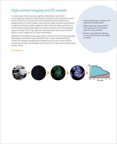
High-Content Imaging and 3D Models
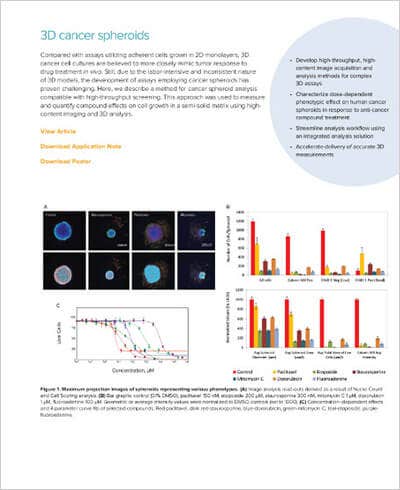
3D Cancer Spheroids
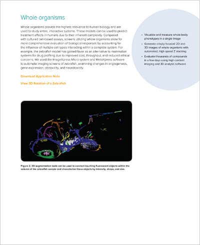
Whole Organisms
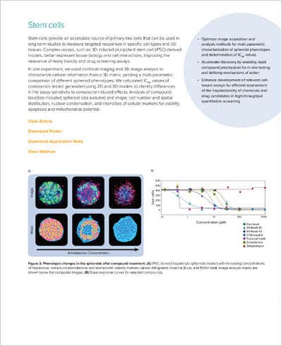
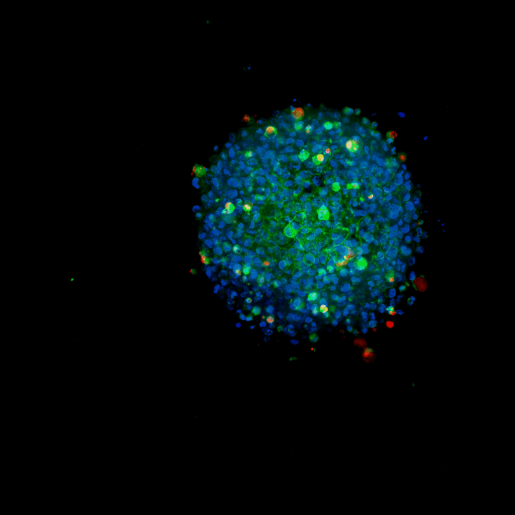
Stem Cells
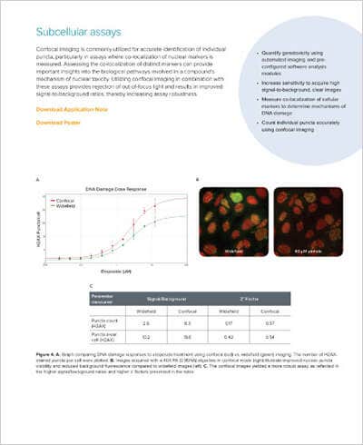
Subcellular Assays
EXPLORE 3D CELL MODEL APPLICATIONS
Register to download your free eBook today

Whatever your imaging requirement, we have a solution
that will meet the needs of your lab and your budget.
3D Cellular Imaging Systems
Our ImageXpress portfolio of cellular imaging systems and analysis software offers innovative capabilities for evaluating complex and emerging 3D cell culture-based models such as:
- Organoids
- Spheroids
- Whole organisms
- Induced Pluripotent Stem Cell (iPSC)-derived cell models
- Organ-on-a-chip technologies
Check your email, your material is on the way!