
Visikol team develops 3D cell culture models to simulate the NASH and NAFLD disease progression process using ImageXpress Micro Confocal High-Content Imaging System and MetaXpress software
COMPANY/UNIVERSITY
Visikol, Inc.
TEAM MEMBERS
Megan Casco, Ph.D., Senior Scientist
Tom Villani, Ph.D., Founder and CSO
Peter Worthington, Ph.D., Director of Drug Discovery
Katie Worley, Ph.D., Principal Scientist
PRODUCTS USED
ImageXpress Micro Confocal High-Content Imaging System
MetaXpress High-Content Image Acquisition and Analysis Software
The Challenge
In the Non-Alcoholic Steatohepatitis (NASH) and Nonalcoholic Fatty Liver Disease (NAFLD) space, there has been a strong need for an in vitro model that allows researchers to better screen out potential therapeutics during lower cost and medium throughput in vitro assays. Liver disease affects millions of people in the United States annually, yet there are few effective therapeutic treatments. Visikol is focused on shifting the paradigm of how tissues and cells are analyzed from a qualitative human-driven approach to a quantitative computer vision driven approach. They offer services to pharmaceutical and biotech clients to help them accelerate their drug discovery efforts and to reduce the translational gap between in vitro and in vivo studies through the use of advanced cell culture models and analysis techniques.
The early stages of both NASH and NAFLD are known to be reversible but researchers have struggled to develop treatments for reversing this process. One of the main difficulties in developing these treatments is that there have not been predictive in vitro models that accurately represent the liver and the NASH or NAFLD disease progression in vitro. Because of this lack of in vitro models, there have been several late-stage clinical trial failures and the cost and time to develop these treatments has been significant.
In addition, imaging 3D cell culture models is challenging as biological material scatters light effectively and image quality deteriorates rapidly in thicker specimens. Further, processing three-dimensional data sets can be difficult to achieve uniform 3D tissue labeling.
The Solution
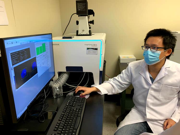
For imaging, Visikol used the ImageXpress® Micro Confocal High-Content Imaging System which allows researchers to fully visualize 3D cell culture models – or spheroids – in their entirety when combined with Visikol® HISTO-M™. The MetaXpress® High-Content Image Acquisition and Analysis Software provided a layout of each well in the plate (each well with one spheroid). The Visikol team selected the correct wavelength of light to excite the fluorophores in the model which emitted a different wavelength of light that was captured by the imager camera. Manual thresholding followed, allowing for the identification of positively stained collagen regions. Typically, imaging is done with 20 Z-slices which allows the camera to capture the entire range of the spheroid which is on average 150 to 250 microns thick. Compressing the Z-slices using the max intensity emission from each slice allowed for the creation of the Z-stack which was a compilation of the brightest emissions from each slice in one image. The resulting images provided visualization of multiple immunolabeled channels including Albumin, Vimentin, CD68, CD31, and DAPI.
Imaging the models allowed researchers to visualize how compounds interact with the cells which gives an understanding of how a compound could affect people in clinical trials. The important aspect of 3D models to NASH is the analysis of scar tissue that forms in the model. Imaging can also reveal the viability of the cells in the model in relation to the client’s compounds.
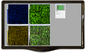
MetaXpress High-Content Image Acquisition and Analysis Software
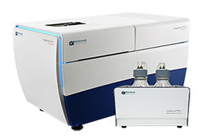
ImageXpress Micro Confocal High-Content Imaging System
Products Used
The ImageXpress Micro Confocal system is a high-content solution that can switch between widefield and confocal imaging of fixed and live cells. It can capture high quality images of whole organisms, thick tissues, 2D and 3D models, and cellular or intracellular events. The spinning disc confocal and sCMOS camera enable imaging of fast and rare events like cardiac cell beating and stem cell differentiation. With the MetaXpress software, the system enables many confocal imaging applications from 3D assay development to screening.
The Results
The team at Visikol have developed several 3D cell culture models mimicking the tissue microenvironment within the liver to simulate the NASH and NAFLD disease progression process. These models are evaluated using 3D confocal high content imaging combined with other readouts and various therapeutic treatments to quantify to what degree they improve the fibrosis process associated with NASH.
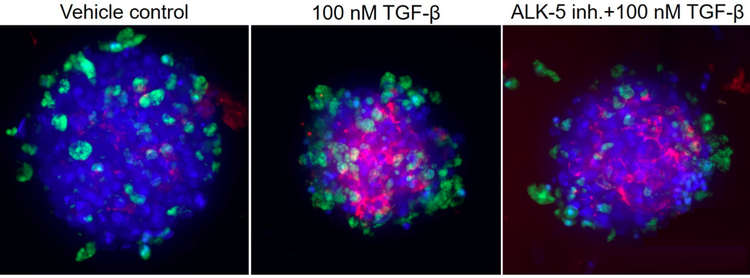
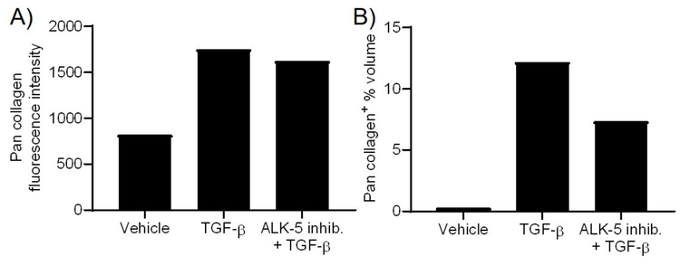
Visikol HepaRG and non-parenchymal co-culture 3D spheroids were either pretreated with vehicle or an ALK-5 inhibitor for 1 h prior to inducing fibrosis with 100 nM TGF-β for 48 h. In the above images, DAPI is in blue, viability indicator is in green and pan collagen is in red. Pan collagen fluorescence intensity as well as volume were quantified for each 3D cell culture model as described below. It can be seen that the TGF-β reduces cell viability as well as the size of the spheroid and also initiates collagen deposition whereas the ALK-5 inhibitor is able to partially ameliorate both reductions in cell viability and collagen deposition.