
Water Immersion Objectives
What is water immersion technology?

Water immersion, in terms of high-content imaging, is the technology that automatically and consistently places a layer of water between the objective (magnification) and the sample.
Many cell imaging systems are designed to have air between the objective and the sample. When using air objectives for 3D imaging, a significant portion of the fluorescent light that is emitted from a sample is refracted away from the objective and becomes lost. This reduces the signal that is collected from the sample, and causes the sample to appear distorted.
Oil objectives have been the gold standard for 3D confocal imaging, because they collect more light from the sample, which increases the signal intensity and enhances the image resolution. However, oil objectives are still problematic, as oil can be difficult to handle in an automated fashion and these objectives can also cause some distortion when imaging large 3D samples.
Water immersion objectives for the ImageXpress Micro Confocal system
Scientists often experience difficulties screening complex models and obtaining better data quality. With water immersion objectives on the ImageXpress® Micro Confocal High-Content Imaging System, they can increase signal up to four times, improve z-resolution, and decrease optical aberrations for sharper, crisper images.
To address 3D image analysis workflow challenges, our MetaXpress® 3D analysis software module enables scientists to generate greater phenotypic data within a single interface without compromising throughput or data quality, giving scientists confidence in their discoveries.

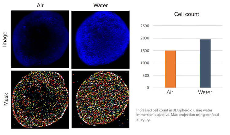
Increased average intensity in 3D assay using water immersion objective. Nuclei of spheroids at 50 ms exposure, maximum projection using confocal imaging.
†Data and images were acquired during development using customer samples. Results may vary.
Gain the sensitivity to capture more phenotypic data at greater depths
Water immersion objectives can offer signal increases of up to 4X† which can help scientists see deeper into 3D and thick tissue samples at lower exposure times.

Gain up to 4 times more signal at depth
Improve z-resolution and decrease optical aberrations for more accurate reconstruction of 3D samples.

Gain sharper, crisper images
Better light collection results in images with brighter intensity at lower exposure times, contributing to better quality of data.

Run your samples with confidence
MetaXpress software includes integrated, user-intuitive sensors and alerts to indicate the system health of the water controller system to give you peace of mind when running your samples.

Acquire 3D samples at high throughput
The ImageXpress Micro Confocal system with water immersion objectives automatically replenishes the water during acquisition, so the entire microplate sample can be acquired at high throughput.

Simplify viewing and quantification of 3D structures
Analyze spheroids, microtissues, cells in a 3D matrix, and small organisms that are acquired as a stack of z-planes. Utilize the MetaXpress 3D Analysis Module to evaluate volume, XYZ location, distance to neighboring objects, diameter, depth, various intensity measurements, texture, or number of objects.

Shorten image acquisition time
QuickID targeted acquisition images at low magnification to find objects of interest or rare events, then, automatically acquires high-magnification images with the required wavelengths, z-stack, or time points. This analysis process shortens acquisition time and reduces imaging storage requirements by acquiring selected targets at high-magnification.
†Data and images were acquired during development using customer samples. Results may vary.