Solid state lasers for high-performance laser excitation
With the increasing use of highly-complex cell-based 2D and 3D assays in biologic research, there is a pressing demand for improvements to the capabilities of automated high-content imaging. The ImageXpress® Confocal HT.ai High-Content Imaging System is a high-throughput solution that utilizes an 8-channel high-intensity laser light source to penetrate into deep tissue while reducing exposure times and increasing focus.
The illumination power of the laser light source significantly increases image intensity and assay sensitivity, which is especially important for dim samples. Imaging systems with lasers substantially decrease exposure times, which result in increased imaging speed and assay throughput. 3D imaging especially benefit from the laser light source with higher image intensities and improve image quality, which result in increased assay sensitivity and imaging speed.
**Data and images were acquired during development using customer samples. Results may vary. Highlighted features’ price, time to deliver, and specifications will vary based on mutually agreed technical requirements. Solution requirements may cause adjustment to standard performance.
AgileOptix technolgy with powerful, solid state light engine
AgileOptix technology makes it possible for the system to deliver the sensitivity and throughput needed for demanding applications by combining an advanced solid-state light engine with seven laser lines and eight filter combinations, proprietary spinning disk technology, state-of-the-art optics, and a scientific CMOS sensor.
Molecular Devices can successfully tailor the ImageXpress Confocal system to include customized software and hardware including the features described herein, as well as integration of other lab components such as incubators, liquid handlers, and robotics for a fully automated work cell. Check out our new Organoid Innovation Center where we showcase these cutting-edge technologies with novel 3D biology methods to address key challenges of scaling complex 3D biology.
Sales are subject to our Custom Product Purchase Terms available at www.moleculardevices.com/custom-products-purchase-terms.
Laser and LED light source specifications
The new modification of the ImageXpress Confocal HT.ai system equipped with multiline laser light source with matched filters spanning 405 nm to 730 nm excitation wavelength provides drastically increased illumination power to the sample. Laser light source results in brighter images, increased sensitivity, and increased speed and throughput speed of assays. The impact is especially important for the assays where sensitivity and timing are limiting factors.
Decreased exposure times with laser illumination
Confocal imaging and 3D image analysis are especially useful for capturing the complexity of 3D biological assays like organoids and spheroids. Here, we compared a standard imaging system with LED excitation to one with laser excitation.
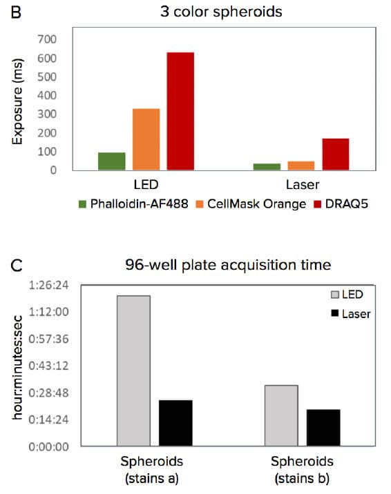
Imaging 3D cancer spheroids

A. DRAQ5 and Phalloidin-AF488 overlay of an untreated spheroid plus nuclear segmentation and area masks of the 2D projection image. Low magnification QC of spheroid staining could be used to locate treated and control spheroids in the well for accurate centering before acquiring at a higher magnification. B. Exposure times optimized to acquire images of equivalent brightness. Different exposures were required for different staining protocols. C. Speed of acquiring a 96-well spheroid plate in the ImageXpress Confocal HT.ai system at 20X using LED vs laser light sources.

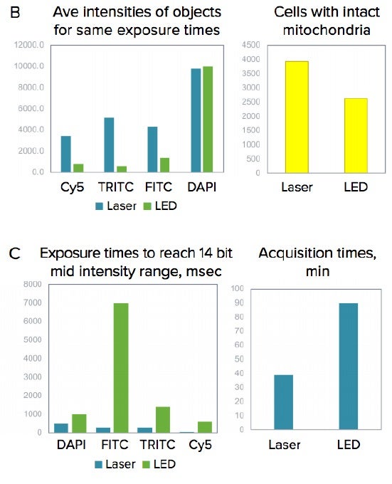
Imaging 3D lung organoids

A. Confocal image of organoid culture (maximum projection), 10X. B. Images were taken using same exposure times for LED or laser light sources, then cell count and average intensities of objects (cells) shown for representative images. C. Exposure times were matched for 14-bit intensity range for both laser and LED. Optimized exposure times and duration of the experiment were substantially reduced by using lasers.

-
Organoid Innovation Center
Our lab automation solutions include scientists and engineers who can customize our instruments, as well as automate entire workflows to meet the specific needs of your assay, method, or protocol. From incubators, liquid handlers, and robotics to customized software and hardware—and with over 35 years of experience in the life science industry—you can count on us to deliver quality products and provide worldwide support.
Learn more about how robotics-driven automation workcells and AI-based image analysis can help you develop an efficient, end-to-end workflow for your organoid development process.
3D Cell Models
3D cell cultures offer the advantage of closely recapitulating aspects of human tissues including the architecture, cell organization, cell-cell and cell-matrix interactions, and more physiologically-relevant diffusion characteristics. Utilization of 3D cellular assays adds value to research and screening campaigns, spanning the translational gap between 2D cell cultures and whole-animal models. By reproducing important parameters of the in vivo environment, 3D models can provide unique insight into the behavior of stem cells and developing tissues in vitro.
-
Improve sensitivity, speed, and assay quality
Improve sensitivity, speed, and assay quality for complex biological assays
Using a high-power laser light source, the system significantly increases light throughput to the sample, which results in brighter images, increased sensitivity, and increased assay throughput. The impact is especially important for the assays where sensitivity and imaging time are the limiting factors.
Monitor lung organoids using 3D high content imaging
Organoid models have increasingly gained popularity in biologic research and screening to recapitulate complexity of real tissues. To model the in vivo human lungs, we have cultured primary human lung epithelial cells under conditions that promote the formation of 3D structures recapitulating the morphological and functional characteristics of the airway.
-
Organoids
Organoids are three-dimensional (3D) multi-cellular microtissues that are designed to closely mimic the complex structure and functionality of human organs. Organoids typically consist of a co-culture of cells which demonstrate a high order of self-assembly to allow for an even better representation of complex in vivo cell responses and interactions, as compared to traditional 2D cell cultures.
Using a laser light source for high-content automated imaging
In this study, we demonstrate improvement in assay sensitivity, precision, and speed of acquisition with a new configuration of the ImageXpress® Confocal HT.ai High-Content Imaging System that utilizes laser light source. The system drastically increases the power of illumination that results in brighter images, increased sensitivity, and increased speed and throughput speed of assays.
Latest Resources
Resources for Confocal Laser Illumination
Flyer
Custom Organoid Expansion Service
Molecular Devices has global leadership in the scale-up and industrial manufacture of human-derived 3Dorganoids using our proprietary bioprocess. Organoids offer significant benefits ov…
Blog
Advanced technology for automated 3D biology workflows #SLASEurope2022
SLAS Europe 2022, hosted numerous sessions packed with the latest research on emerging topics as well as sessions and panel discussions focused on how to build, and succeed, in a…
Blog
How automated organoid cell cultures are developed, imaged, and analyzed
3D cell models are becoming increasingly popular for studying complex biological effects, tissue functionality, and diseases. Their ability to self-organize and mimic…
Blog
Engineering Next-gen Organoids with Automated Lab Workflows at #SLAS2022
SLAS2022, the Society for Lab Automation and Screening conference offered another exciting year for learning about innovative laboratory technologies. Whether you attended in-person…
Blog
Stem cell science insights and breakthroughs presented at #ISSCR2021
If you didn't get a chance to visit us at our poster sessions during ISSCR 2021, don’t fret. We've gathered all our sessions right here for you. The ISSCR Annual Meeting brought…
Publications
Project Profile: Organoid Innovation Center
Lab Manager speaks to Dan O’Connor, vice president, drug discovery, Molecular Devices, about the company’s Organoid Innovation Center in San Jose, CA. The center is 180 sq. ft., within…
Publications
Organs-on-Chips: Expand the Boundaries of In Vitro Testing
Humans began culturing animal cells and tissues as early as the late 19th century, when Wilhelm Roux first showed that chick embryos could be cultured in saline solution for a few days.…
Blog
Tips for running a successful live cell imaging experiment
There have been significant advancements in microscopy and camera technology, as well as advancements in technologies for labeling molecules of interest over the past decade. These…
Blog
Overcome the challenges of high-throughput 3D imaging
Thanks to recent advances in imaging technologies, we are now able to observe and analyze complex cellular networks in three dimensions. Through 3D imaging, we can acquire and…
Publications
Shaping the Future of Organoid Research
Molecular Devices, a provider of innovative life science technology, recently unveiled a brand new, first-of-its-kind Organoid Innovation Center. Situated at the company’s global headqu…
Publications
How AI is advancing neurodegenerative disease research
In this guest editorial, explore how artificial intelligence is becoming a powerful tool for drug discovery As part of the SelectScience Advances in Drug Discovery Special Feature, gues…
Brochure
ImageXpress Confocal HT.ai
The ImageXpress® Confocal HT.ai High-Content Imaging System utilizes a seven-channel laser light source with eight imaging channels to enable highly multi-plexed assays while maintainin…
Publications
Using 3D cancer cell models to push forward personalized medicine
Using 3D cancer cell models to push forward personalized medicine: an interview with Angeline Lim, PhD, of Molecular Devices Editor-in-Chief, Francesca Lake, speaks to Angeline Lim of…
Publications
Danaher’s SCIEX and Molecular Devices Businesses Debut New Products
It has been a busy beginning of the year for Danaher’s Life Sciences segment. In January, the business reported 2020 revenues of $10.6 billion, representing 47% of the total revenues of…
Scientific Poster
Using a laser light source for high-content automated imaging
In this study, we demonstrate improvement in assay sensitivity, precision, and speed of acquisition with a new configuration of the ImageXpress® Confocal HT.ai High-Content Imaging Syst…
Application Note
Improve sensitivity, speed, and assay quality for complex biological assays
Here, we demonstrate improvement in assay sensitivity, quality, and speed of acquisition with the ImageXpress® Confocal HT.ai, our high-content imaging laser-based system.
Blog
Water immersion technology and high-content imaging: A closer look
Three-dimensional (3D) cellular assays have emerged as a valuable tool in drug discovery and biological research, as they closely mimic in vivo environments and are shown to…
Publications
Advanced Cell Screening and Imaging Reveals How the Cardiac System Responds in Drug Development
In drug discovery, time and monetary investments increase significantly the further candidates advance through the preclinical phase of testing, analysis and evaluation. By excluding no…
Publications
Improving acquisition and analysis of 3D cell model assays with water immersion objectives
Sebastian Peck is a senior product manager for cellular imaging at Molecular Devices, one of the world's leading providers of high-performance bioanalytical measurement systems, softwar…
Publications
The workflow game-changer: combined technologies for efficiency
Microfluidic flowchip technology combined with three-dimensional (3D) imaging is a powerful duo poised to change the future of drug discovery and development. The integration of combine…
Publications
Imaging system produces human cellular morphological data for COVID-19 research
In an effort to better understand the cellular responses to COVID-19, the digital biology company Recursion has publicly released the world’s largest imaging dataset portraying therapeu…
Publications
3D Microscopy Keeps Getting Faster, Smarter, Leaner
With each passing day, continuous advancements are being made in the studies that cutting-edge instruments can perform and in the complexity of the biological samples that are being exa…
Publications
From 3D high throughput to simple automated imaging: Cellular imaging within reach of every laboratory
OUR ABILITY to image cells has come a long way since the pioneering days of Galileo Galilei and Antonie van Leeuwenhoek. The multiple imaging techniques available today range from simpl…
Flyer
High-performance Customizations
Capture more data at greater depths for 3D and thick tissue samples Scalable, high-performance, high-content screening solutions with the ImageXpress® Micro Confocal system
Videos & Webinars

Enhancing 3D Disease Models: Automated, High-Throughput, Phenotypic Screening with Organ-on-a-Chip