AgileOptix spinning disk confocal microscopy
Our proprietary AgileOptix™ Spinning Disk Technology is a microscopy innovation designed to run more complex, physiologically-relevant cellular models including 3D spheroids, tissues, live-cell assays, and whole organisms. This technology combines the clarity of confocal imaging with the throughput and well-known robustness of the ImageXpress® Confocal systems.
Unlike other spinning disk confocal technologies used for high-content screening, AgileOptix technology offers the ability to adapt disk geometries to meet your unique assay requirements. It is one of the few technologies on the market that includes a unique dual disk configuration with both pinholes or slits to accommodate high-throughput applications.


Multiple confocal disk configurations for optimized assay performance
AgileOptix Spinning Disk Technology includes simple, user-defined configurations to ensure maximum assay performance for both confocal and widefield imaging. Multiple confocal disk geometries are available including pinhole and slit configurations.
Spinning disk geometry | 60 µm pinhole (single disk) |
60 µm dual disk with 50 µm slit |
60 µm dual disk with 42 µm pinhole |
High-sensitivity detection |
|
|
|
Fast acquisition |
|
|
|
>3 log dynamic range† |
|
|
|
Widefield mode for flat biology |
|
|
|
Most confocal applications |
|
|
|
Highest resolution imaging |
|
||
High-throughput applications |
|
*Powered by our highly responsive sCMOS sensor and advanced solid state light source.
How spinning disk confocal technology works
One aspect of AgileOptix technology is a spinning disk light path, which includes a very bright solid state light engine. Excitation light passes through a rapidly spinning disk with symmetrically placed spirals of pinholes or slits. These pinholes or slits split the illumination light into multiple beams that scan the fluorescent sample. Emission light from the sample then passes back through the confocal disk and is then directed via a dichroic beamsplitter through the emission filter to a high-sensitivity scientific CMOS camera.
Optimize out-of-focus light or “haze” that can skew image analysis
AgileOptix spinning disk technology optimized for high resolution, tissue penetrance, and high throughput
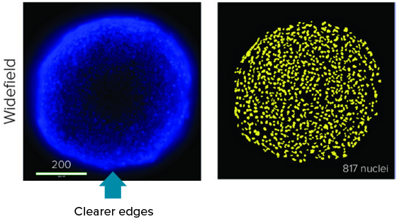
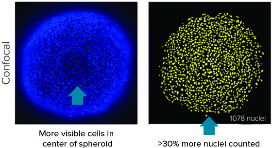

Deep tissue penetrating, confocal disk module
Specialized deep tissue penetrating, confocal disk module, combined with a laser light source, improves light penetrance for deeper tissue penetration, resulting in sharper images with improved resolution when imaging thick tissue samples†.
- Improve suppression of out-of-focus light
- Reduce haze (pinhole crosstalk)
- Penetrate deeper into thick tissue samples for sharper images
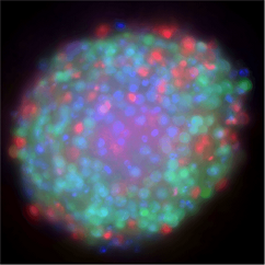
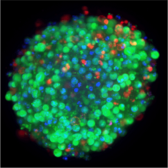

Images taken at the same exposure

**Data and images were acquired during development using customer samples. Results may vary. Highlighted features’ price, time to deliver, and specifications will vary based on mutually agreed technical requirements. Solution requirements may cause adjustment to standard performance.
Molecular Devices can successfully tailor the ImageXpress Confocal system to include customized software and hardware including the features described herein, as well as integration of other lab components such as incubators, liquid handlers, and robotics for a fully automated work cell. Check out our new Organoid Innovation Center where we showcase these cutting-edge technologies with novel 3D biology methods to address key challenges of scaling complex 3D biology.
Sale is subject to our Custom Product Purchase Terms available at www.moleculardevices.com/custom-products-purchase-terms.
More great resources
-
Organoid Innovation Center
Our lab automation solutions include scientists and engineers who can customize our instruments, as well as automate entire workflows to meet the specific needs of your assay, method, or protocol. From incubators, liquid handlers, and robotics to customized software and hardware—and with over 35 years of experience in the life science industry—you can count on us to deliver quality products and provide worldwide support.
Learn more about how robotics-driven automation workcells and AI-based image analysis can help you develop an efficient, end-to-end workflow for your organoid development process.
3D Cell Models
3D cell cultures offer the advantage of closely recapitulating aspects of human tissues including the architecture, cell organization, cell-cell and cell-matrix interactions, and more physiologically-relevant diffusion characteristics. Utilization of 3D cellular assays adds value to research and screening campaigns, spanning the translational gap between 2D cell cultures and whole-animal models. By reproducing important parameters of the in vivo environment, 3D models can provide unique insight into the behavior of stem cells and developing tissues in vitro.
-
Improve sensitivity, speed, and assay quality
Improve sensitivity, speed, and assay quality for complex biological assays
Using a high-power laser light source, the system significantly increases light throughput to the sample, which results in brighter images, increased sensitivity, and increased assay throughput. The impact is especially important for the assays where sensitivity and imaging time are the limiting factors.
Organoids
Organoids are three-dimensional (3D) multi-cellular microtissues that are designed to closely mimic the complex structure and functionality of human organs. Organoids typically consist of a co-culture of cells which demonstrate a high order of self-assembly to allow for an even better representation of complex in vivo cell responses and interactions, as compared to traditional 2D cell cultures.
Latest Resources
Resources for AgileOptix Spinning Disk Confocal Technology
Blog
3D Biology: The paradigm shift in next-generation drug discovery [Interactive infographic]
The drug discovery landscape is changing. Bringing a drug to market has historically been time-consuming and costly, with many candidates failing in the first phase of clinical trials…
Application Note
CloneSelect Imager FL fluorescence for rapid day zero monoclonality
Gene editing allows DNA manipulation, i.e., addition, deletion, or modification using different tools. Several nucleases for genome editing are now available: zinc finger nucleases (…
Scientific Poster
Structural organization in 3D human iPSC-derived cardiac tri-culture microtissues
The human heart is a complex organ providing highly regulated processes of moving blood through the body. The adult human ventricle is comprised of cardiomyocytes, endothelial cells, fi…
Blog
How automated organoid cell cultures are developed, imaged, and analyzed
3D cell models are becoming increasingly popular for studying complex biological effects, tissue functionality, and diseases. Their ability to self-organize and mimic…
Scientific Poster
Automation of the organ-on a chip assay: automated culture, imaging and analysis of angiogenesis
There is a critical need for biological model systems that better resemble human biology. Three-dimensional (3D) cell models and organ-on-a-chip (OoC) structures representing various ti…
Blog
Engineering Next-gen Organoids with Automated Lab Workflows at #SLAS2022
SLAS2022, the Society for Lab Automation and Screening conference offered another exciting year for learning about innovative laboratory technologies. Whether you attended in-person…
Publications
Project Profile: Organoid Innovation Center
Lab Manager speaks to Dan O’Connor, vice president, drug discovery, Molecular Devices, about the company’s Organoid Innovation Center in San Jose, CA. The center is 180 sq. ft., within…
Publications
Organs-on-Chips: Expand the Boundaries of In Vitro Testing
Humans began culturing animal cells and tissues as early as the late 19th century, when Wilhelm Roux first showed that chick embryos could be cultured in saline solution for a few days.…
Blog
Tips for running a successful live cell imaging experiment
There have been significant advancements in microscopy and camera technology, as well as advancements in technologies for labeling molecules of interest over the past decade. These…
Blog
Overcome the challenges of high-throughput 3D imaging
Thanks to recent advances in imaging technologies, we are now able to observe and analyze complex cellular networks in three dimensions. Through 3D imaging, we can acquire and…
Publications
Shaping the Future of Organoid Research
Molecular Devices, a provider of innovative life science technology, recently unveiled a brand new, first-of-its-kind Organoid Innovation Center. Situated at the company’s global headqu…
Publications
How AI is advancing neurodegenerative disease research
In this guest editorial, explore how artificial intelligence is becoming a powerful tool for drug discovery As part of the SelectScience Advances in Drug Discovery Special Feature, gues…
Publications
Using 3D cancer cell models to push forward personalized medicine
Using 3D cancer cell models to push forward personalized medicine: an interview with Angeline Lim, PhD, of Molecular Devices Editor-in-Chief, Francesca Lake, speaks to Angeline Lim of…
Publications
Danaher’s SCIEX and Molecular Devices Businesses Debut New Products
It has been a busy beginning of the year for Danaher’s Life Sciences segment. In January, the business reported 2020 revenues of $10.6 billion, representing 47% of the total revenues of…
Blog
Water immersion technology and high-content imaging: A closer look
Three-dimensional (3D) cellular assays have emerged as a valuable tool in drug discovery and biological research, as they closely mimic in vivo environments and are shown to…
Publications
Advanced Cell Screening and Imaging Reveals How the Cardiac System Responds in Drug Development
In drug discovery, time and monetary investments increase significantly the further candidates advance through the preclinical phase of testing, analysis and evaluation. By excluding no…
Publications
Improving acquisition and analysis of 3D cell model assays with water immersion objectives
Sebastian Peck is a senior product manager for cellular imaging at Molecular Devices, one of the world's leading providers of high-performance bioanalytical measurement systems, softwar…
Publications
The workflow game-changer: combined technologies for efficiency
Microfluidic flowchip technology combined with three-dimensional (3D) imaging is a powerful duo poised to change the future of drug discovery and development. The integration of combine…
Publications
Imaging system produces human cellular morphological data for COVID-19 research
In an effort to better understand the cellular responses to COVID-19, the digital biology company Recursion has publicly released the world’s largest imaging dataset portraying therapeu…
Publications
3D Microscopy Keeps Getting Faster, Smarter, Leaner
With each passing day, continuous advancements are being made in the studies that cutting-edge instruments can perform and in the complexity of the biological samples that are being exa…
Publications
From 3D high throughput to simple automated imaging: Cellular imaging within reach of every laboratory
OUR ABILITY to image cells has come a long way since the pioneering days of Galileo Galilei and Antonie van Leeuwenhoek. The multiple imaging techniques available today range from simpl…
Flyer
High-performance Customizations
Capture more data at greater depths for 3D and thick tissue samples Scalable, high-performance, high-content screening solutions with the ImageXpress® Micro Confocal system
Brochure
Imaging Solutions to Answer Your Biological Questions
Learn about our imaging family and which solution is best suited to answer your biological questions.
Videos & Webinars

Automation of the organ-on-a-chip assay: automated culture, imaging and analysis of angiogenesis

Challenges for drug screening of complex biological systems

3D Tissue Models Imaging and Automation of Organ-On-A-Chip

SLAS Dublin 2022

Introduction to the Organoid Innovation Center and ImageXpress Confocal HT.ai system

Gain deeper insights into cellular 3D structures with water immersion objectives