Advancements in Early Toxicity Testing [Podcast]
Toxicity is one of the most pervasive pain points in drug development. Approximately 90% of drug candidates fail in clinical trials, 30% of which are caused by unmanageable toxicity1, even though each drug is developed over 10-15 years with significant investment and resources. Many previously approved drugs are withdrawn from the market due to their adverse effects on cardiac, liver, and renal cells, neurons, or blood cells. This brings to question the validity of animal models used for toxicological testing of drugs. Animal models often fail to predict toxicity in humans due to their different genomic makeups, metabolism, and life spans. Therefore, replacing traditional animal studies with more predictive toxicity assessment methods has become a necessity.
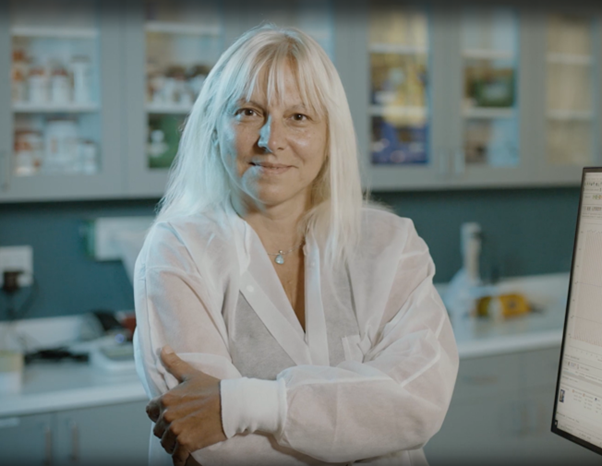
In the new episode of the Drug Target Reviews podcast, Dr. Ruth Roberts, co-director and co-founder of ApconiX, and Dr. Oksana Sirenko, senior scientist at Molecular Devices, discuss how advanced 3D cell models can improve in vitro toxicity testing. They also discuss the role of high-throughput screening and emerging machine-learning technologies in improving the speed and reliability of toxicological studies.


Key learning points:
Why is early toxicity detection crucial?
The unprecedented toxicity of a drug is a serious setback for patients and drug innovators. Withdrawals or terminations caused by adverse effects result in delayed delivery to patients in critical need. It can also harm patients participating in the clinical trials. Thus, detecting potential toxic effects early in drug development pipelines helps companies to derisk complications.
- Dr. Ruth Roberts
Since many drugs require billions of dollars of investment and years of development, toxicity can be detrimental to company resources. Dr. Roberts points to the impact toxicity detection in later stages can have on pharmaceutical companies. "When we lose a drug due to toxicity later on in the drug development process, we also waste significant investment of financial resources and significant amount time, not to mention the animals sacrificed for this purpose". Therefore, it is essential to generate models for toxicological testing early in drug development for timely delivery to patients and for enhancing patient quality of life.
How advanced cell models and organoids transform toxicity testing
Toxicity test methodologies started to move away from animal models in the search for more predictive and reliable models. In that regard, 3D cell models, such as spheroids, organoids, and organ-on-a-chip systems, can represent human body conditions more accurately than 2D in vitro cell lines and animal models. In particular, organoids are ideal platforms to generate human-like conditions due to the morphological organization of different cell types and the interactions between them.
Dr. Sirenko believes that organoids offer several benefits over traditional models in measuring toxicity. "3D cell cultures may present dose-dependent responses, much like in human tissues and organs. Scientists can measure the impact of a drug on morphological characteristics like neuronal growth or angiogenesis. The sensitivity of 3D models enables the detection of toxicity more quickly and at lower concentrations through functional activity assessment, as opposed to cell viability assays."
https://share.vidyard.com/watch/cxAetNR6yCyXn4xjYQBmca
Cardiac organoids derived from induced pluripotent stem cells (iPSCs) are widely used for functional assays for drug toxicity screening in Molecular Devices' applications. These organoids can mimic cardiac muscle contractions, which can be visualized using calcium-sensitive fluorescent dyes and recorded using imaging instruments, thus indicating drug cardiotoxicity. Changes to functional characteristics occur in case of drug-induced toxicity. For cardiac organoids, changes to contractions and ion channel activity are used as robust indicators of potential toxic effects. Liver organoids are also susceptible to enhanced toxicity, mimicking various responses occurring in the human body, such as apoptosis, steatosis, and autophagy, as well as altered secretion of analytes, such as alkaline phosphatase or lactate dehydrogenase.
These positive outcomes have started to encourage the FDA to welcome non-animal methodologies to evaluate drug safety. While animal models are still the mainstay, continued improvement of 3D cell models can accompany and eventually replace animal testing.
The contribution of high-throughput screening to toxicity testing?
High-throughput screening is a critical milestone in drug discovery, as it allows the evaluation of large numbers of drug candidates for efficacy and safety. Researchers can work through diverse drug chemistry and diverse targets to find a group of optimum drug-target conditions.

This workflow helps illustrate an integrative in vitro assay using human iPSC-derived cardiomyocytes for the high-throughput screening of several diverse classes of environmental chemicals and drugs (i.e. NTP screening library). Learn more about this study, In Vitro Cardiotoxicity Assessment of Environmental Chemicals Using an Organotypic Human Induced Pluripotent Stem Cell-Derived Model.
An added benefit, according to Dr. Sirenko, is the ability to isolate more drug candidates in a given time. "Traditionally, drug development funnel involves eliminating drug candidates in each step because of the cost and the hardship of carrying on multiple drug candidates later in a development step. Using high-throughput screening on advanced cell models early in drug development greatly helps to determine the number of candidates with or without toxicity from early stages. So, you will not have to eliminate toxic candidates later in development." Overall, high-throughput screening significantly increases productivity and speed in drug development workflows.
The importance of machine learning and AI in toxicity assessment
Manipulation of increasingly complex data requires powerful analytical methods. Machine learning algorithms help researchers deduce patterns in complex data and provide insights into drug mechanisms of action. Recent breakthroughs in artificial intelligence (AI) can facilitate advances in medicine and healthcare, including diagnosis, patient stratification, development of personalized medicine, and toxicity assessment.
Advanced drug discovery process often involves high throughput image acquisition of 3D biological models treated with a panel of compounds, followed by automated analysis of those images. Certain compounds may alter the morphology of the biological models, causing unwanted toxic effects, and therefore, it is important to detect such perturbations in a robust and reliable manner. One of the key applications of AI in drug discovery is image and data analysis. To support researchers, Molecular Devices offers IN Carta Image Analysis Software, which has an intuitive interface while leveraging state-of-the-art AI capabilities for complex assays. It allows deep learning models to be trained to detect multicellular organoids, cells, or subcellular structures in the images of 3D biological models. Once the AI model is trained, IN Carta can help discern whether any given compound within a panel has a positive effect, exhibits toxicity, or has no effect compared to the control treatments.

Classification of spheroids formed from HCT116 cells. Spheroids were segmented based on brightfield images using SINAP. Sample was additionally counter-stained with with Hoechst 33342, Calcein AM and MitoTracker Red to visualize nuclei, live cells and mitochondria respectively.
Besides image analysis, there are several other uses of ML/AI in toxicity testing. In fact, Dr. Roberts has participated in a multitude of projects at ApconiX. One of these projects involves employing the Toxicogenomic Generative Adversarial Network (TGCAN) machine learning tool that predicts toxicogenomic profiles of cell models upon drug treatment. "We have trained the model on toxicogenomic profiles from rat liver with a multitude of compounds and doses. Thus, we could predict genomic profiles for compounds that have not been through testing". Additionally, she participated in other projects that implement deep learning tools to extract and collate essential information from complex drug safety assessment reports, which can reduce the time needed to glean insight into drug toxicity.
Tissue-Specific Toxicity Testing For High-Throughput Drug Discovery
Early in vitro toxicity testing is vital for the timely detection of potential adverse effects a drug can exert. Molecular Devices offers cutting-edge 3D cell models derived from human cells to generate toxicity profiles that are more congruent with human biology than what 2D cell models can offer. Furthermore, we offer comprehensive toxicity workflows comprising a multitude of assays to assess cell viability and functionality. These include MTT assays, cell titer glow assays, plate reader assays, imaging methods, live-dead assays, and apoptosis assays, which can assess the potentially toxic effects of drugs on cardiac cells, hepatocytes, and neuronal cells. These workflows can also integrate high-throughput screening and advanced AI tools for rapid and accurate analysis of toxicity tests. For researchers focused on high-throughput cellular screening of cardiomyocytes, neurons, and other cell models, the FLIPR® Penta High-Throughput Cellular Screening System provides real-time kinetic assays for early toxicity assessment. Learn more about the FLIPR Penta system here.
- Sun, Duxin, et al. "Why 90% of clinical drug development fails and how to improve it?." Acta Pharmaceutica Sinica B 12.7 (2022): 3049-3062.