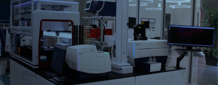
Organoid Innovation Center
Modular, automated 3D cell culture and image analysis lab enables customers to streamline and scale organoid interrogation, advancing drug discovery research
Automated 3D cell culture and image analysis lab streamlines and scales complex biology research
The new Organoid Innovation Center at Molecular Devices combines cutting-edge technologies with novel 3D biology methods to address key challenges of scaling complex 3D biology.
The collaborative space brings customers and researchers into the lab to test automated workflows for organoid culturing and screening, with guidance from in-house scientists.
An end-to-end solution standardizes the organoid development process with cell culture, treatment, and incubation, through to imaging, analysis, and data processing, delivering consistent, unbiased, and biologically-relevant results at scale.
Quickly adopt innovative, 3D biological methods and technologies for drug discovery
The center expands beyond imaging to demonstrate a fully-integrated solution that addresses the challenges associated with every step in the sample prep-to-report pipeline for assays performed on complex 3D biological models.
The Organoid Innovation Center showcases cutting-edge instruments that work harmoniously together for autonomous, long-term, live cell 2D and 3D cell culture growth and monitoring with intelligent label-free imaging. This integrated workflow provides quality control alerts and readiness, 3D organoid screening, and deep learning image analysis that reveals hidden patterns other technologies miss.
See the power and flexibility of an automated and customizable high-throughput screening solution
With intuitive scheduling software researchers can control the 3D workflow remotely, tracking the cell journey from single cell to differentiated organoid, along the way. Cell culture and incubation is streamlined with an automated incubator and collaborative robot that maintains culture consistency. Media exchange for culture maintenance is standardized and streamlined with automated liquid handling, minimizing manual intervention. 3D model development can be monitored over time with label-free imaging to assess assay readiness – and with real-time feedback, scheduling of automated compound addition and treatment is standardized.
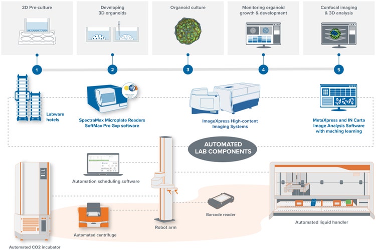
Organoid Screening Workflow
- Step 1) 2d Pre-culture – Organoids are pre-cultured from iPSC-derived or primary cells (intestine, lung, or brain)
- Step 2) Developing 3D Organoids – Cells are transferred to 24-well plates then placed in an incubator to promote cell growth and differentiation into specific tissue in 3D
- Step 3) Organoid Culture – Organoid culture process require multiple steps with different media exchanges
- Step 4) Monitor organoid growth & development – Organoid are monitored and characterized for complex analysis of tissue structure and differentiation
- Step 5) Confocal imaging and 3D analysis – The visualization and characterization of multiple quantitative descriptors used for studying disease phenotypes and compound effects
Our new ImageXpress Confocal HT.ai High-Content Imaging system is designed for 3D imaging. This system offers eight high-powered laser excitation channels and automated water immersion objectives that boost signal and assay sensitivity without sacrificing speed. Spinning disk confocal technology with five pinhole geometry options reduces haze from out-of-focus light for deeper organoid penetration and improved axial resolution. For analysis of complex 3D biology, IN Carta Image Analysis Software provides a streamlined workflow with powerful deep-learning-based segmentation, machine learning-based classification, and 3D volumetric analysis.
Danaher Launches Collaboration with Cincinnati Children’s Hospital Medical Center Aiming to Improve Patient Safety in Early Drug Development
Molecular Devices Introduces CellXpress.ai Automated Cell Culture System, a 3D Biology Innovation Hub for Fast, Reliable Drug Discoveries
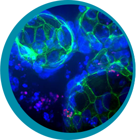
Molecular Devices launches custom organoid line expansion service using unique bioprocess technology for high-throughput applications

Molecular Devices inks deal with HUB Organoids to advance automated intestinal organoid screening technology
Complete solutions for a fully integrated lab automation workflow
Our lab automation solutions include scientists and engineers who can customize our instruments, as well as automate entire workflows to meet the specific needs of your assay, method, or protocol. From incubators, liquid handlers, and robotics to customized software and hardware—and with over 35 years of experience in the life science industry—you can count on us to deliver quality products and provide worldwide support.
Sale is subject to our Custom Product Purchase Terms available at www.moleculardevices.com/custom-products-purchase-terms
Molecular Devices is honored to be designated as a "Lab of the Future" Company
Exhibitors that received the SLAS2024 Lab of the Future designation have demonstrated their ability to provide solutions that go beyond automated instrumentation alone to push the boundaries of current technology and break new ground in achieving complete integration of automated workflows in the lab space.
Take a step ‘back to the future’ and revisit us at SLAS 2024 – explore six posters that feature this groundbreaking innovation and unveil the latest advancements in laboratory research and technology.
Lung organoid cell image gallery
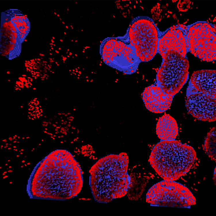
Image analysis of organoid
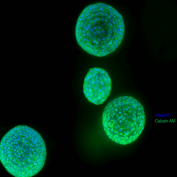
Lung organoids
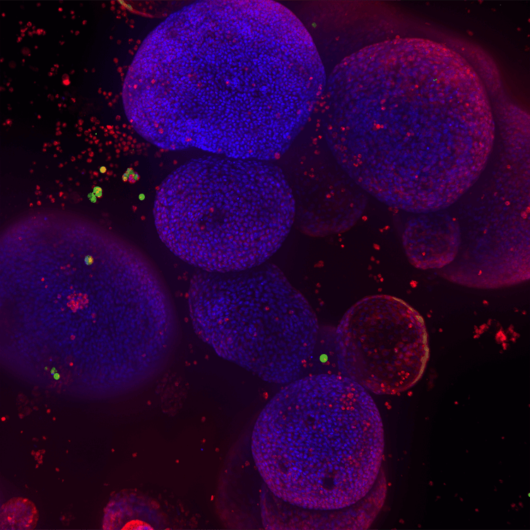
Lung organoids 2
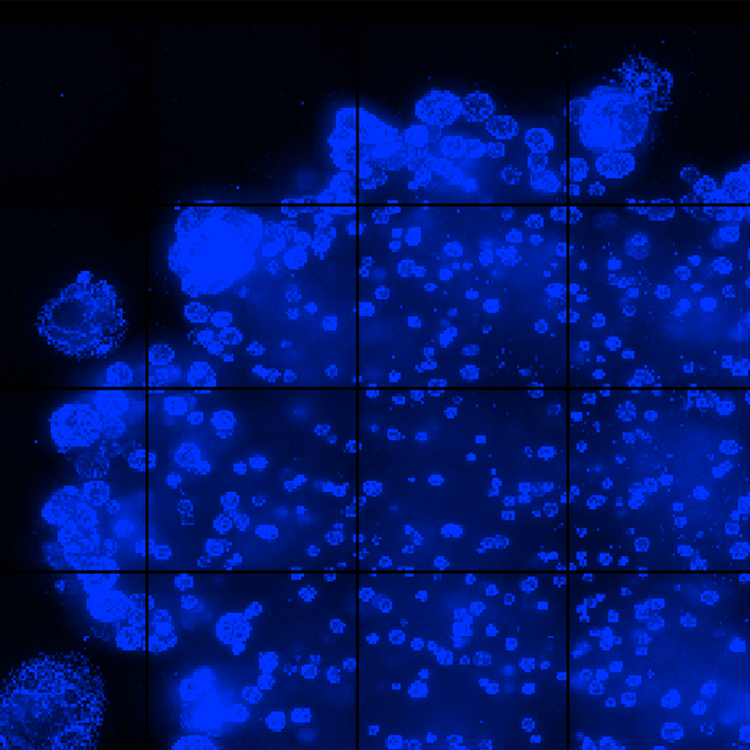
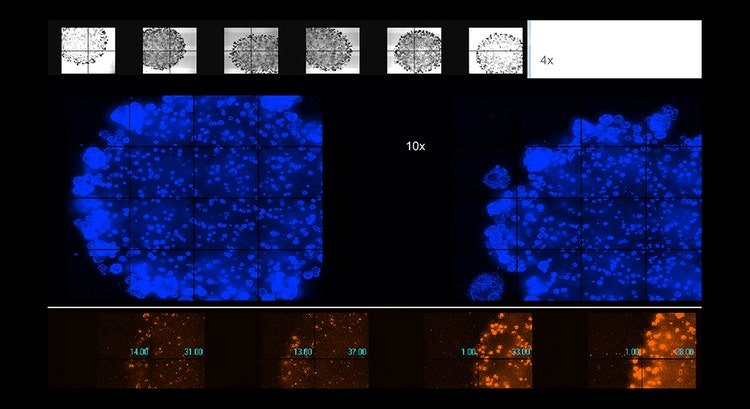
Automated imaging of live organoids in Matrigel was done using confocal option, 4x or 10x magnification
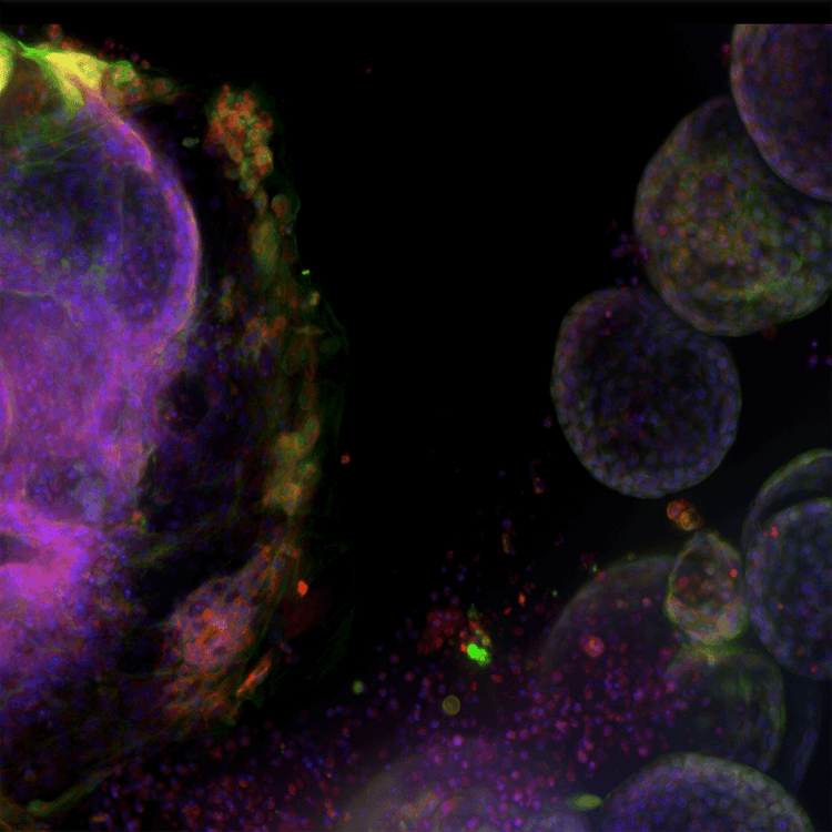

High complexity lung organoid
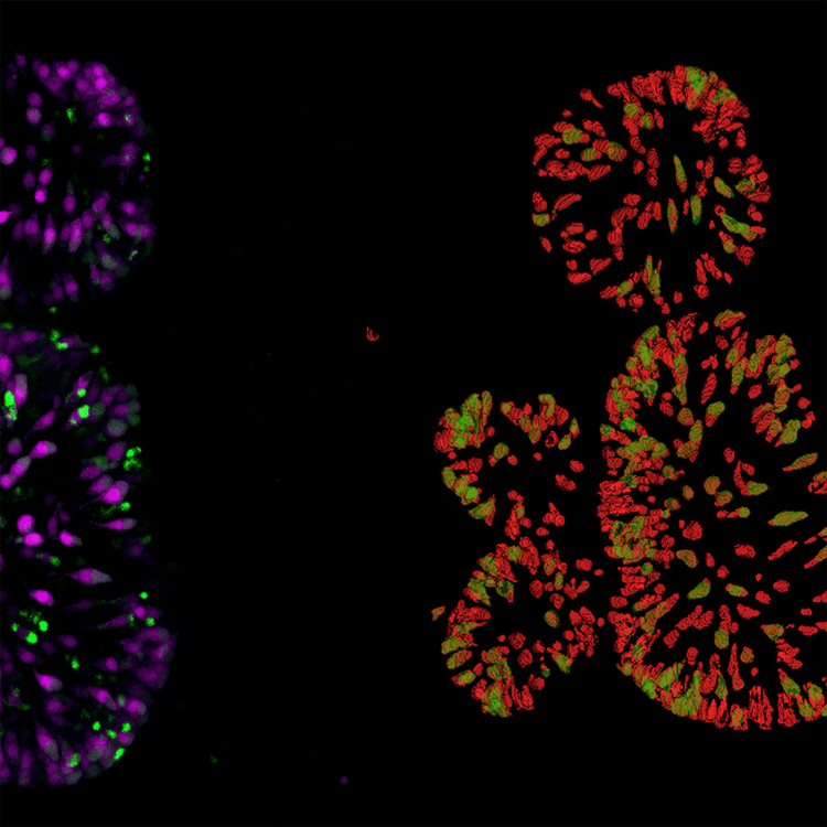
Gut organoid dTomato cells in red Detection of Double positive(mNeon) in green
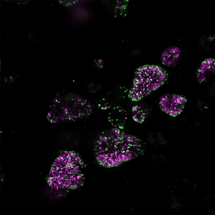
Internally expressed genes dTomato magenta mNeon green 10x
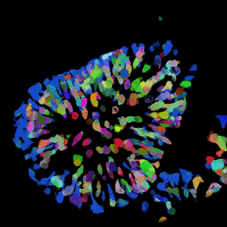
Intestinal organoid 40x,dTomato cell count
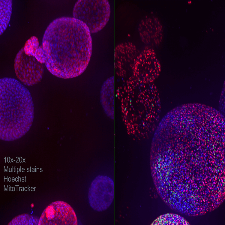
Lung organoid 10x 20x Multiple stains Hoechst MitoTracker
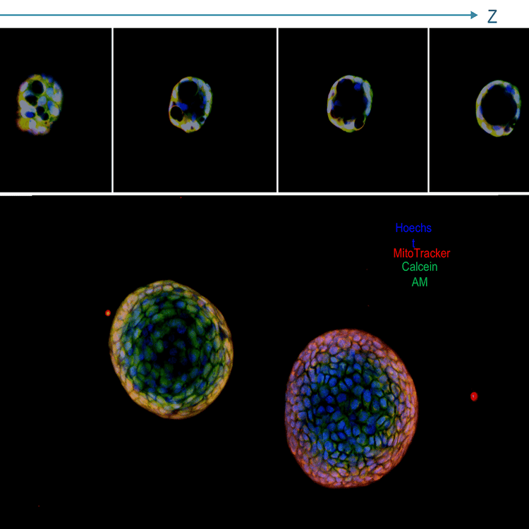
Lung organoids,projection image and a panel with Z stack; live stained with Hoechstblue,nuclei),MitoTracker(red,mitochondria),and Calcein AM(green,viability dye);20x water immersion
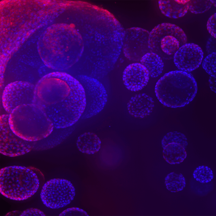
Lung organoids 3
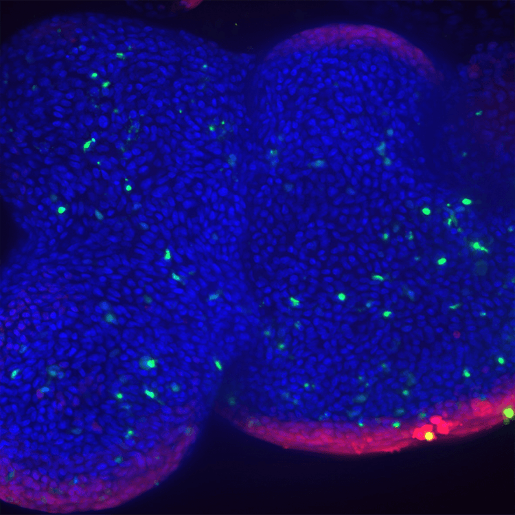
Lung organoids 4
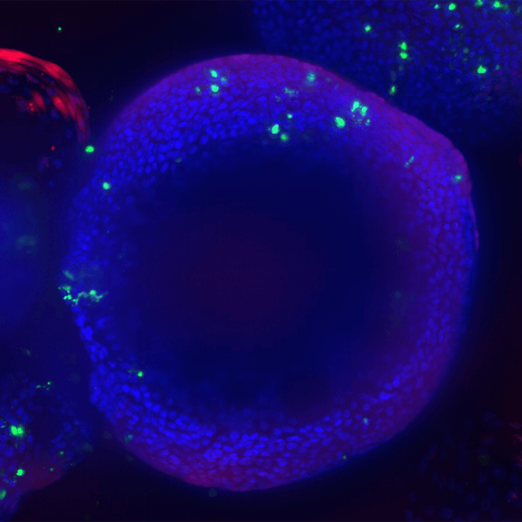
Lung organoids lung airway epithelial cells cultured in matrigel domes for 8 weeks
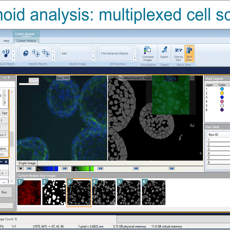
Organoid analysis multiplexed cell scoring
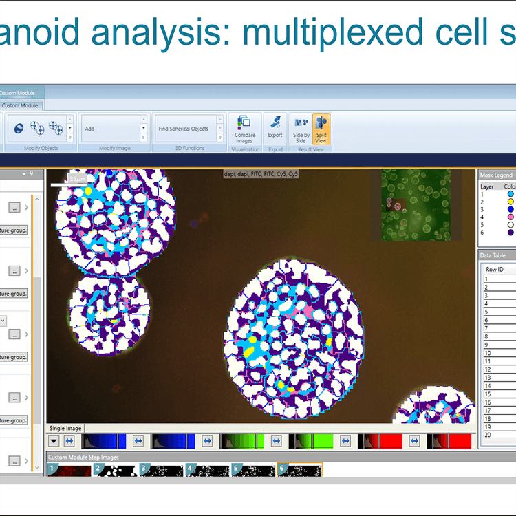
Organoid analysis multiplexed cell scoring 2
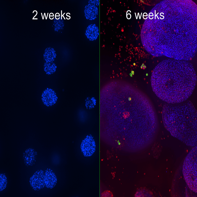
Organoid development
Applications and assays
The Organoid Innovation Center builds on Molecular Devices 35 years of experience delivering high-performance life science technology to customers for improved drug development, biotechnology research, and clone screening workflows.
Learn more about our industry leading 3D biology and automated high-content imaging applications: