TRF, TR-FRET & HTRF

Download eBook

TRF / TR-FRET (HTRF) Overview

Time-Resolved Fluorescence (TRF)

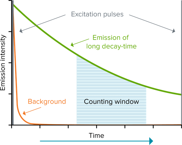
Fluorescence intensity (FI) measurements use standard fluorophores like fluorescein, whose emission is short-lived, on the order of nanoseconds. Excitation of the sample and measurement of emission occur simultaneously. Although microplate readers are very good at screening out excitation light from the emission measurement, that excitation light, along with short-lived light emitted by materials in the well or sample, often contributes to high background.

Time-resolved fluorescence (TRF) reduces background by using a lanthanide fluorophore, such as europium or terbium, that emits long-lived fluorescence. This long-lived fluorescence lasts for milliseconds, so excitation of the fluorophore by a pulsed light source (e.g., a flash lamp), followed by a delay and then signal measurement (‘counting window’), allows short-lived fluorescence (lasting only for nanoseconds) to subside before a measurement is made. Assays using time-resolved fluorescence offer dramatically increased signal-to-noise ratios. The most frequently used lanthanides are europium, terbium and samarium. These are commonly used as chelate or cryptate complexes that enable good signal intensity and stability.


​​​​​​Time-Resolved Fluorescence (TRF)

Time-Resolved Fluorescence Resonance Energy Transfer (TR-FRET)


Time-Resolved Fluorescence Resonance Energy Transfer (TR-FRET)​​​​​

no FRET

Donor and acceptor are distant

FRET

Donor and acceptor are inclose proximity

TR-FRET combines the time-resolved (TR) measurement of fluorescence with fluorescence resonance energy transfer (FRET) technology. In FRET assays, biomolecules (e.g., proteins) are labeled with donor and acceptor fluorophores. When the biomolecules interact, donor and acceptor fluorophores are brought close together. Now, when the donor is excited, it can transfer its emission energy to the acceptor, which in turn emits fluorescence at a specific wavelength. Acceptor and donor fluorescence emissions have different wavelengths that can be distinguished from each other by a microplate reader, enabling quantitation of the biomolecular interaction.

Using lanthanide fluorophores, which have long-lived fluorescence emission, as donors, TR-FRET assays take advantage of the time-resolved measurement of fluorescence to eliminate short-lived background fluorescence. In a TR-FRET assay, thanks to the donor fluorophore’s long-lived emission, excitation and emission of both donor and acceptor fluorophores can also be measured after short-lived background fluorescence has abated.

Homogeneous Time-Resolved Fluorescence (HTRF)

Homogeneous Time-Resolved Fluorescence (HTRF)​​​​​

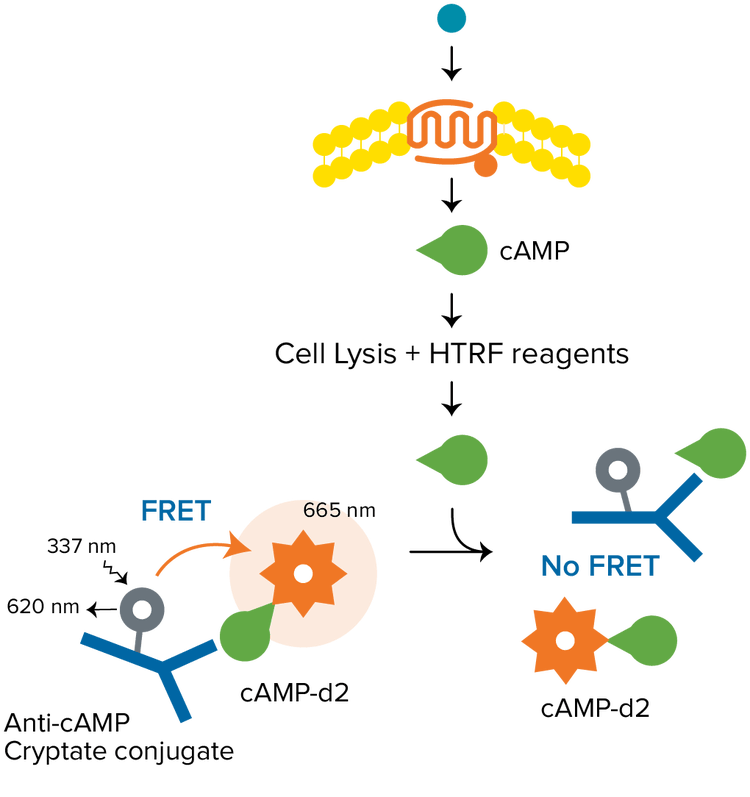
HTRF is a versatile TR-FRET technology developed by Cisbio for detecting biomolecular interactions. A typical HTRF assay uses a europium cryptate as the donor, and the organic fluorophore d2 as the acceptor. The donor and acceptor can be used to label a variety of biomolecules, for applications including epigenetics, biomarker quantification, GPCR signaling, and much more. HTRF assays require a microplate reader with TRF detection mode that is certified HTRF-compatible by Cisbio.

Customer Breakthroughs

iTeos uses our multi-mode microplate readers to identify therapeutics that target the immune tumor micro-environment

Learn more  


Advantages and Considerations

A key benefit of TRF and TR-FRET detection includes decreased background and increased signal-to-noise ratio compared to standard fluorescence, resulting in higher sensitivity. In addition, the assay has a robust mix-and-read format that does not require any washing. This, along with the assay’s stability, makes it easy to automate and miniaturize for screening applications.


Applications and Assays

TRF and TR-FRET (HTRF) assays enable the analysis of molecular interactions in biochemical processes and are widely used to study kinase assays, cellular signaling pathways, protein-protein interactions, DNA-protein interactions, cell cytotoxicity and receptor-ligand binding.

Example assays for TR-FRET are outlined in the table to the right, along with several time-resolved fluorescence resources below.
 

Assay Donor Acceptor Wavelengths
HTRF-Eu/red* Europium cryptate XL665 or d2 Ex320 Em1 620/Em2 665
HTRF-Tb/red* Terbium cryptate XL665 or d2 Ex340 Em1 620/Em2 665
HTRF-Tb/green* Terbium cryptate fluorescein or GFP Ex340 Em1 620/Em2 520
Lanthascreen Tb** Terbium chelate fluorescein Ex340 Em1 490/Em2 520
Lanthascreen EU** Europium chelate Alexa Fluor 647 Ex320 Em1 620/Em2 665
LANCE (original)*** Europium chelate Surelight® APC Ex320 Em1 620/Em2 665
LANCE Ultra*** Europium chelate ULight Ex320 Em1 620/Em2 665

​​​​​​

  • Cytokine assays

    HTRF Human TNFα Assay

    Pro- and anti-inflammatory cytokines play a central role in autoimmune, inflammatory and infectious diseases. They are also key players in metabolic disorders and oncology, particularly the anti-tumor immune response. HTRF assays provide a versatile platform for the quantitation of various cytokines and chemokines and are suited to monitoring cytokine and chemokine release in cell-based experiments. With the SpectraMax i3x Multi-Mode Microplate Reader and SpectraMax® MiniMax™ 300 Imaging Cytometer, users are able to measure HTRF and cell viability, as well as confirm assay quality by monitoring cells’ appearance.

    Explore our resources for cytokine assays:

    eBook: From ELISA to epigenetics with HTRF

    ELISA to epigenetics with HTRF

    Overcome HTRF detection challenges with optimized microplate reader settings and simplified analysis.

    eBook: From ELISA to epigenetics with HTRF

  • HTRF cAMP assays, Gαi/o-coupled receptors

    HTRF cAMP dynamic 2 and IP-One assays

    Cyclic AMP (cAMP, cyclic adenosine 3’, 5’–monophosphate) is a key second messenger in GPCR signaling. The generation of cAMP is probably the most studied of signal transduction pathways and is involved in responses to sensory input, hormones, nerve transmission, and much more. Upon ligand binding to a GPCR, a conformational change occurs, activating the receptor and in turn activating a G protein. Activation of Gαi/o–coupled receptors inhibits adenyl cyclase from generating cAMP.

    Explore our resources for Gαi/o

    HTRF cAMP assays, Gαs-coupled receptors

    HTRF cAMP assays

    Cyclic AMP (cAMP, cyclic adenosine 3’, 5’–monophosphate) is a key second messenger in GPCR signaling. The generation of cAMP is probably the most studied of signal transduction pathways and is involved in responses to sensory input, hormones, nerve transmission, and much more. Upon ligand binding to a GPCR, a conformational change occurs, activating the receptor and in turn activating a G protein. Further signal transduction depends on the type of G protein activated, but activation of Gαs leads to upregulation of cAMP by adenylate cyclase.

    Explore our resources for Gαs

  • HTRF assay

    HTRF assay

    HTRF assays from Cisbio have evolved to encompass the areas of G protein-coupled receptors, kinases and cell signaling, epigenetics, and biomarkers. A wide variety of toolbox reagents and kits are available to address protein-protein interactions, nuclear receptor assays, receptor dimerization, ligand binding, receptor internalization, enzyme assays, and many more.

    HTRF involves the use of two fluorophores, a donor and an acceptor, to label proteins or other biomolecules of interest. When the biomolecules interact, their proximity enables fluorescence resonance energy transfer (FRET) to occur from the donor to the acceptor fluorophore. Using long-lifetime fluorescent lanthanide cryptates as donors allows time-resolved detection of HTRF assays, minimizing short-lived background fluorescence from compounds and other materials. The detection of time-resolved FRET (TR-FRET) with a microplate reader provides a readout of biomolecular binding.

    HTRF-compatible microplate readers

    HTRF-compatible microplate readers

    Getting the best HTRF results requires a microplate reader that has been validated to meet the HTRF performance specifications set by Cisbio. A reader’s optics, energy source, and detectors, as well as the settings used to detect HTRF, all impact performance. Our readers use optimized components for HTRF detection, including an HTRF detection cartridge for the SpectraMax i3x and Paradigm Multi-Mode Microplate Readers, and an enhanced TRF module plus validated filters for the SpectraMax iD5 reader.

    Cisbio’s HTRF certification program guarantees that readers bearing the “HTRF compatible” logo meet the specifications required for optimal HTRF performance. This technical note presents the optimized settings validated by Cisbio for Molecular Devices HTRF-compatible microplate readers.

  • IP-One assays, Gαq/11-coupled  receptors

    IP-One assays, Gαq/11-coupled receptors

    Phospholipase C (PLC) is another common target of activated G proteins and is critical to a variety of processes. For example, thrombin receptors in platelets use this pathway to promote blood clotting. Cisbio IP-One competitive immunoassays detect the accumulation of inositol monophosphate (IP1), a stable downstream metabolite of IP3 induced by the stimulation of PLC following Gαq/11-coupled receptor activation. This kit allows direct characterization of receptor agonists and antagonists, in both adherent and suspension cells.

    Kinase & epigenetic assays

    Kinase & epigenetic assays

    Protein kinases are enzymes that play a central role in various signal transduction pathways involved in the control of cell growth, metabolism, differentiation, and apoptosis. Kinases are the second most important group of drug targets after GPCRs. Epigenetics is the study of heritable phenotype changes that do not involve alterations in nucleotide sequences, for example, changes such as DNA methylation and histone modification. These have been linked with the development of various diseases, particularly in cancer.

    Explore our resources for kinase & epigenetic assays:

  • Measure oxidative metabolism and glycolytic activity

    Measure oxidative metabolism and glycolytic activity

    To survive, cells require fuel in the form of ATP to carry out most essential bioprocesses. This fuel is generated through glycolysis and mitochondrial respiration. While both produce ATP, glycolysis can function in the absence of oxygen, whereas mitochondria require oxygen for the final step in oxidative phosphorylation (OXPHOS). Understanding how these pathways respond to effector compounds can provide useful insights into the overall function of cells and the underlying mechanisms that determine cell fate.

    Read our application note to learn about real-time data acquisition and analysis of the progression of oxygen consumption and glycolic activity:

    Optimized settings for Transcreener TR-FRET assays

    Transcreener TR-FRET assays

    The Transcreener® TR-FRET Assays are single step, competitive immunoassays for direct detection of nucleotides with a far-red time resolved Förster resonance energy transfer (TR-FRET) readout. Learn how to optimize settings for Transcreener TR-FRET assays on the SpectraMax iD5 and i3x multi-mode microplate readers.

Latest Resources

Resources of Time-Resolved Fluorescence TRF / TR-FRET (HTRF)

Videos & Webinars

How to move from time-consuming processes to efficient workflows with lab automation solutions

How to move from time-consuming processes to efficient workflows with lab automation solutions

SpectraMax Mini

SpectraMax Mini