Fluorescence Polarization (FP)
Fluorescence polarization (FP) is a technique that is widely used to monitor binding events in solution. It can be used to assess biomolecular interactions, including protein-antibody binding and DNA hybridization, as well as enzyme activity, and it has been adapted to basic research as well as high-throughput screening.
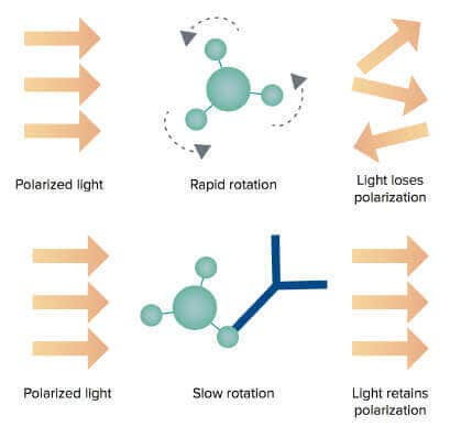
A small, fluorescently labeled molecule (tracer) that is excited with plane-polarized light emits mostly depolarized light because the tracer tumbles rapidly during the time between excitation and emission. However, when the tracer binds a much larger molecule, it rotates more slowly, and the emitted light remains largely polarized.
Fluorescence polarization G factor
Polarization, expressed in units of milli P, or mP, is calculated from the measurements of perpendicular (Iperp) and parallel (Ipara) fluorescence intensity values detected relative to the direction of the polarized excitation light (see the formula below). The G factor (G) is used to correct for the effects of optical components like filters, polarizers, and monochromators, which can affect polarization values.
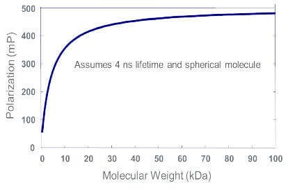
The larger the fluorescently labeled molecule, or the molecule to which tracer is bound, the larger the mP value.
FP advantages over other binding assays
FP can be employed to study receptor-ligand interactions, protein-DNA interactions, proteolysis, membrane fluidity, enzyme assays, and more. FP assays have been used successfully to investigate a wide variety of targets including kinases, phosphatases, proteases, G protein–coupled receptors (GPCRs), and nuclear receptors.
The following benefits make FP assays especially amenable to high-throughput screening:
- Homogeneous (no wash steps required)
- Non-radioactive
- Ratiometric (single wavelength)
- Miniaturizable
IMAP phosphodiesterase assays on SpectraMax Multi-Mode Microplate Readers
IMAP® Technology from Molecular Devices enables rapid, homogeneous, and non-radioactive assay of kinases, phosphatases, and phosphodiesterases and is suited for both assay development and high-throughput screening. IMAP assays are based on binding of phosphate to immobilized metal coordination complexes on nanoparticles. When IMAP binding entities bind to phosphorylated substrate, molecular motion of the peptide is altered, and fluorescence polarization (FP) for the fluorescent label attached to the peptide increases (Figure 1, left). In a TR-FRET version of the assay, the inclusion of a Terbium (Tb) donor enables a fluorescent energy transfer to occur when phosphorylated substrate is present (Figure 1, right). This assay is detected in a time-resolved mode, which virtually eliminates fluorescence interference from assay components or compounds in a screen. TR-FRET also offers flexibility in substrate size and concentration.
Cyclic nucleotide phosphodiesterases (PDEs) are a group of enzymes that degrade the phosphodiester bond of cAMP and cGMP, second messengers that are involved in a variety of biological processes. They have emerged as a key class of druggable targets due to their clinical significance in areas including heart disease, dementia, depression, and others. Here, we demonstrate how PDE enzyme dilution and inhibition curves are performed with IMAP Technology using the SpectraMax® Multi-Mode Microplate Readers with SoftMax® Pro Software.
Figure 1: A phosphodiesterase reaction is performed using a fluorescent-labeled substrate. Binding Solution containing large M(III)-based nanoparticles is then added. In the FP readout (left), the small phosphorylated fluorescent substrate binds to the large nanoparticles, which reduces the rotational speed of the substrate and thus increases its fluorescence polarization. In the TR-FRET readout (right), phosphorylated substrate and Tb donor both bind to the nanoparticle, bringing the Tb donor in close proximity to the fluorescein acceptor on the substrate and enabling FRET.
Streamline the workflow for measuring IgG in cell line development
The measurement of IgG production is a crucial step at many stages in the development and manufacturing of monoclonal antibodies. Commonly used methods for IgG quantitation require either special instrumentation and skilled personnel, such as HPLC and surface interferometry, or time-consuming assays like ELISA (enzyme-linked immunosorbent assay). Although the ELISA is a well-established method for protein quantitation, it is a lengthy, multi-step process. Here, we introduce the use of the Valita®TITER assay from Valitacell for measuring IgG titers during the antibody development and manufacturing process.
The ValitaTITER assay is based on detection of IgG Fc interactions with Protein G using fluorescence polarization (FP) technology. Each well of a 96- well microtiter plate is coated with a fluorescently labeled IgG-binding peptide, Protein G. When samples are added to the wells, the Protein G molecules are resuspended and binding occurs. The rate of molecular motion of Protein G slows down when it is bound to antibodies, resulting in an increase of the FP value (Figure 2).
Figure 2: Free Protein G molecules are smaller and rotate faster in the buffer solution. As a result, the polarized excitation light loses polarization (top). When Protein G molecules bind to the antibodies in the samples, the rotation of the larger complex slows down resulting in increase of the FP values (bottom).
More great resources for fluorescence polarization assays
-
Establishing and optimizing a fluorescence polarization assay
This technical note is designed to provide information to help the user define the optimal experimental conditions for converting existing assays to a robust fluorescence polarization format. The example is a competitive binding assay of the type often used to evaluate receptor-ligand binding. Familiarity with the basic principles of fluorescence polarization is assumed.
IMAP FP kinase assays
Protein kinases are central to the regulation of many cellular processes. In recent years they have emerged as one of the most important classes of drug targets for cancer and many other diseases. IMAP® Technology from Molecular Devices enables rapid, non-radioactive assay of a wide array of kinases and is suited to both assay development and high-throughput screening.
-
IMAP phosphodiesterase assay
IMAP Technology offers a homogeneous, non-antibody-based platform for screening kinases, phosphatases, and phosphodiesterases. Stable and robust, it is amenable to miniaturization and yields high-quality data with excellent Z’ factor values, making it suitable for inhibitor screening. Unlike some other methods, IMAP measures direct binding for more relevant results. IMAP TR-FRET assays also enable direct Km determination.
IMAP technology
Until now, assays of kinase activity have been performed using radioactive isotopes or highly specific antibodies. To address this, Molecular Devices introduced its proprietary IMAP® Technology, providing a non-radioactive, homogeneous assay applicable to a wide variety of kinases without regard for the substrate peptide sequences. The assay is a simple “mix-and-read” procedure that allows accurate determination of enzyme activity
-
Transcreener ADP2 assays
Transcreener® ADP2 Assays are homogenous assays with fluorescent readouts that enable the detection and screening of established drug targets including protein and lipid kinases, as well as emerging targets such as carbohydrate kinases, triphosphatases, heat shock proteins and other ATPases. The assay is based on the immunodetection of ADP. Three detection modes are oered to accommodate users’ needs and detection format preferences: fluorescence polarization (FP), time-resolved Forster-resonance energy transfer (TR-FRET), and fluorescence intensity (FI).
ValitaTITER assay
The ValitaTITER assay is a homogeneous, high-throughput method for precise and rapid quantitation of IgG in the cell line development workflow. This 96-well assay has been fully validated on the SpectraMax iD5 reader and other Molecular Devices plate readers with FP detection to ensure reliable results. SoftMax Pro Software minimizes setup time for detection and automates standard curve fitting and sample quantitation
-
IgG Quantification
In therapeutic protein engineering and cell line development, IgG quantification is the process of measuring the amount of immunoglobulin G (IgG), a common class of therapeutic proteins, produced by a genetically modified cell line. This is important for evaluating and monitoring the productivity of the cell line and selecting the best candidate for further development of therapeutic antibodies.
Latest Resources
Resources of Fluorescence Polarization (FP)
Publications
The Best Microplate Readers According to our Product Reviewers
According to Biocompare, “one of the most overlooked pieces of equipment in any lab is the microplate reader. While they play an important role in generating data from a wide range of a…
Flyer
Quickly and easily quantify IgG with ValitaTiter
Integrating ValitaTiter and Molecular Devices fluorescence polarization (FP) plate readers produces a seamless IgG titer assay setup and optimization. Cell line development teams will r…
eBook
Virology and Vaccine Research Microplate Reader Solutions
We understand what a challenging time this is for those who are working to get accelerated testing and medicines to the market as soon as possible and are committed to supporting scient…
eBook
The Ultimate Guide to Microplate Reader Solutions
Evaluating microplate readers doesn’t have to be overwhelming. First, consider your application needs. If your budget is modest, a single-mode reader dedicated to your main application…
Publications
Multiple Steps from Microplate-Reader Workstations
Scientists take many roads to overcoming bottlenecks in workflows. For example, a workstation including a microplate reader can handle multiple steps, which improves efficiency and adds…
Application Note
Establishing and optimizing a fluorescence polarization assay
This technical note is designed to provide information to help the user define the optimal experimental conditions for converting existing assays to a robust fluorescence polarization…
Scientific Poster
SpectraMax Microplate Readers : A Complete Solution for Transcreener Assays
Transcreener® ADP2 Assays are homogenous assays with fluorescent readouts that enable the detection and screening of established drug targets including protein and lipid kinases, as wel…
Data Sheet
IMAP Technology for Kinases, Phosphatases and Phosphodiesterases
Get information about proprietary IMAP Technology which provides a non-radioactive, homogeneous assay for kinase activity.
Application Note
IMAP FP kinase assays on the SpectraMax M5 Multi-Mode Microplate Reader
Protein kinases are central to the regulation of many cellular processes. In recent years they have emerged as one of the most important classes of drug targets for cancer and many…
Application Note
IMAP phosphodiesterase assays on SpectraMax Multi-Mode Microplate Readers
Cyclic nucleotide phosphodiesterases (PDEs) are a group of enzymes that degrade the phosphodiester bond of cAMP and cGMP, second messengers that are involved in a variety of biological…
Data Sheet
Fluorescence Polarization SpectraMax Detection Cartridge
Get more information about Fluorescence Polarization SpectraMax Detection Cartridges, which offer great sensitivity for SpectraMax i3x and Paradigm Systems.
Videos & Webinars

How to move from time-consuming processes to efficient workflows with lab automation solutions

Achieve more with the SpectraMax Mini

Expanding Assay Applications with Multi-Mode Readers from ELISA to AlphaScreen