University of Zurich uses the ImageXpress Widefield System to investigate host pathogen interactions
"We needed to be able to image and rapidly quantify fluorescent viral plaque formations accompanied by numerous other markers of choice in an automated and screening-compatible fashion"
– VARDAN ANDRYASIAN
COMPANY/UNIVERSITY
Group of Prof. Dr. Urs Greber, Institute of Molecular Life
Sciences, University of Zurich, Switzerland
TEAM MEMBERS
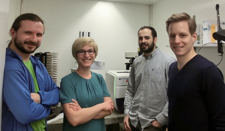
Vardan Andriasyan
Fanny Georgi
Robert Witte
Luca Murer
Dr. Artur Yakimovich (now at MRC Laboratory for Molecular Cell Biology, University College London)
PRODUCTS USED
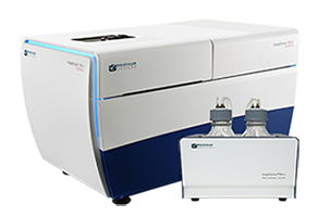
ImageXpress Micro Confocal High-Content Imaging System
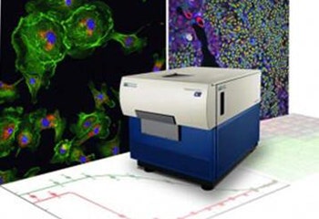
ImageXpress Micro XLS Widefield High-Content Analysis System
The Challenge
At the University of Zurich, researchers are interested in late events that occur in human cells infected by replicating pathogens such as viruses. These events typically involve shedding of the newly synthesized progeny of the pathogen and its spread to non-infected host cells. Their overall aim is to elucidate how viruses take control over membrane and lipid functions and dynamics, cytoplasmic transport processes, and metabolism to support their gene expressions, progeny formation, and ultimately transmission between cells. Research focuses on adenoviruses and rhinoviruses, two agents that cause human respiratory diseases.
One of the first quantitative assays developed in virology was the plaque assay. Originally developed by Renato Dulbecco, it measures the propagation of infectious agents across a monolayer of cells. However, one of the limitations of this assay is the low throughput. The team therefore wanted to enhance this assay by developing a broadly applicable, fluorescence, microscopy-based, high-throughput method to mine patho-biological clonal cell features.

The Solution
To image and analyse infected cells, the group uses an ImageXpress® Confocal system as well as an ImageXpress Widefield system. Using these instruments, together with image analysis software the group developed in house, they have brought the classical plaque assay onto a whole new level - which they call Plaque2.0
The group combines state-of-the-art wet lab, imaging, image and data analysis tools, as well as computer simulations to visualize, measure, and predict the strategies different pathogens exploit to ensure their survival and procreation at the expense of their host.
“What we particularly like about our ImageXpress systems is the combination of flexibility and reliability. They are the workhorses of our group”, says Vardan Andriasyan.

ImageXpress Micro XLS Widefield High-Content Analysis System

ImageXpress Micro Confocal High-Content Imaging System
Products Used
The ImageXpress® Micro Confocal High-Content Imaging System helps you expand the boundaries of your research with the ultimate combination of speed, sensitivity and flexibility. Capture high quality images of whole organism, thick tissues, 3D spheroid assays, and cellular or intracellular events. Combined with MetaXpress® High-Content Image Acquisition and Analysis Software, the ImageXpress Micro Confocal system provides a complete multi-dimensional, high-throughput screening solution to help you discover your next landmark scientific breakthrough.
The ImageXpress Micro XLS High-Content Analysis System has been replaced by the ImageXpress Micro 4 High-Content Analysis System, which represents the culmination of four generations of imaging expertise. The latest agile design allows you to boost your research with this faster-than-ever system, while providing the option to upgrade to confocal in the future to align with your research needs.
The Results
The group’s use of high-content imaging to enhance the classical Plaque assay allows them to image and rapidly quantify viral plaque formations accompanied by numerous other markers of choice in an automated and screening-compatible fashion.
By using the ImageXpress Confocal and Widefield systems to analyze viral-induced cellular events, they can investigate the phenotypic changes that occur during infection. Multi-parametric measurements include infection density, intensity, area, shape or location information at single plaque or population levels. The team employs both live-cell and end-point imaging of cells and viruses and combine this with biochemical analyses and numerical models.
The group is confident that their research will yield new insights into molecular mechanisms underlying cell functions and infection processes and provide a basis for viral applications in clinical research and biotechnology. This may enable the design of new anti-viral agents.
The composite fluorescence microscopy image below (not to scale) depicts the formation of plaques upon infection of cultured cells with adenovirus (blue cells), vaccinia viruses (orange, green) and herpes virus (magenta). The images were acquired using the ImageXpress Micro Widefield System.

References

More information about the Plaque2.0 assay can be found in the following publication:
Yakimovich, A., Andriasyan, V., Witte, R., Wang, I. H., Prasad, V., Suomalainen, M., & Greber, U. F. (2015). Plaque2. 0 - A High-Throughput Analysis Framework to Score Virus-Cell Transmission and Clonal Cell Expansion. PloS one, 10 (9): e0138760.