
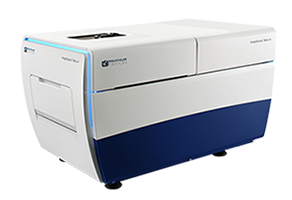
ImageXpress Micro 4 High-Content Imaging System [Discontinued]
This product has been discontinued.
The ImageXpress® Micro 4 High-Content Imaging System is no longer available for purchase. Support for existing customers may continue per applicable service agreements.
For service, documentation, or installed‑base support, please visit SpectraNet.
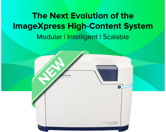
Introducing ImageXpress HCS.ai High-Content Screening System — Modular, intelligent, and built for the future of high-content imaging
Building on the trusted legacy of the ImageXpress Micro 4 system, Molecular Devices introduces the next evolution in high-content imaging — the ImageXpress HCS.ai High-Content Screening System. This fifth-generation modular platform is designed to scale effortlessly with your research, empowering you to move seamlessly from widefield imaging to advanced laser-based confocal modalities—all within one adaptable system.
Engineered for discovery without limits, ImageXpress HCS.ai system combines the renowned optical excellence you trust with revolutionary new capabilities: accelerated acquisition speeds, the intuitive, redesigned MetaXpress® Acquire software, and next-generation, AI-driven IN Carta® analysis to extract deeper insights from complex 2D and 3D models.
Whether you're progressing from widefield microscopy to confocal or expanding into advanced, AI-powered 3D analysis, the ImageXpress HCS.ai system delivers the speed, flexibility, and intelligence to keep your research at the cutting edge—built upon the innovation you already trust.
ImageXpress Micro 4: Widefield imaging for basic research through to high-content screening
The ImageXpress® Micro 4 High-Content Imaging System is a high-throughput, widefield imager that can acquire images of whole organisms and cellular or intracellular events. It features proprietary fast frame rate technology, allowing for the capture of fast biological processes such as calcium oscillations in cardiomyocytes. It is highly configurable and adaptable to changing research needs. Available options include confocal, brightfield, phase contrast, liquid handing, and environmental control.

Support many cellular assays
Image an incredible range of assays with various user-changeable filter cubes, 1-100x objectives, environmental control, fluidics option, and many sample formats including atypical plates for 3D assays.

Analyze images of more applications
A selection of feature-rich turnkey application modules is available with MetaXpress® software. For the development of novel assays, we offer an optional custom analysis module.

Tailor the system to meet your needs
The imager is a highly flexible solution, you can add or change many of the components as your research priorities change. Examples include enhance light engines and automation.
Features

Multiple imaging modes
The system offers fluorescence, widefield, colorimetric, phase contrast, and brightfield label-free imaging.
Upgradeable to confocal
The system can be upgraded to include the proprietary AgileOptix™ technology for accurate 3D volumetric analyses.

Wide dynamic range
Low and high intensity signal quantification in a single image with >3 log dynamic range intensity detection.

High speed image acquisition
Using 4x magnification with a 1536-well plate, 225K wells per day can be acquired. Greater than a million wells per week can be imaged.

Optional environmental control
Temperature, humidity, and CO2 control for multi-day live-cell imaging or fast kinetic studies are available.

Large magnification range
Air and oil immersion objectives ranging from 1x to 100x are available for installation into the 4-position objective changer.
Latest Resources
Featured Applications
Customer Breakthrough
Expand your research using flexible imaging solutions
Molecular Devices provides flexible options for the ImageXpress Micro 4 High-Content Imaging System to meet your research needs and to easily capture images from different sample formats, including hanging drops and in round or flat bottom plates, for monitoring cell health kinetics under environmental control, and more.

ENVIRONMENTAL CONTROL
Environmental control maintains temperature and humidity levels while minimizing evaporation for multi-day, live cell, time-lapse imaging.

TRANSMITTED LIGHT TOWER
Our transmitted light tower enables acquisition of high contrast images for unstained cells which can be easily viewed or separated from background.

ON-BOARD ROBOTIC FLUIDICS
Integrated fluidics automates assay workflows which require compound addition, well washing, and media exchange.
Implement a solution that works for you
Molecular Devices can successfully tailor the ImageXpress Micro 4 High-Content Imaging System to include customized software and hardware including the features described herein, as well as integration of other lab components such as incubators, liquid handlers, and robotics for a fully automated workcell. With over 30 years of experience in the life science industry, you can count on us to deliver quality products and provide worldwide support.
Sale is subject to our Custom Product Purchase Terms available at www.moleculardevices.com/custom-products-purchase-terms
