
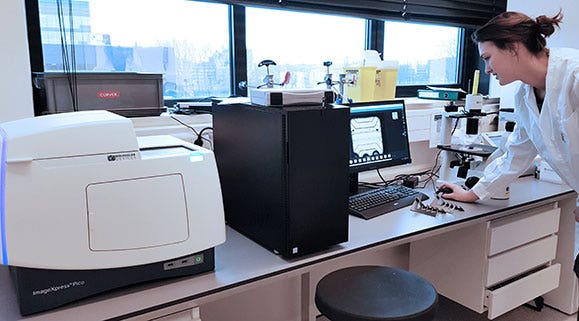
MIMETAS uses the ImageXpress Pico and ImageXpress Micro Confocal systems to develop tissue models for their OrganoPlate®
"The ImageXpress systems enable us to use our 3D organ-on-a-chip platform as a true high-throughput system. The ImageXpress Pico, ImageXpress Micro and ImageXpress Micro Confocal each provide valuable readouts, and all are a perfect match for the OrganoPlate®"
Jos Joore, MIMETAS
COMPANY/UNIVERSITY
MIMETAS
TEAM MEMBERS
Jos Joore, Managing Director and Co-Founder
PRODUCTS USED
ImageXpress Micro Confocal High-Content Imaging System
ImageXpress Micro 4 High-Content Imaging System
ImageXpress Pico Automated Cell Imaging System
The Challenge
MIMETAS offers the OrganoPlate®, a unique 3D organ-on-a-chip platform. The OrganoPlate® is a fully compatible microfluidic culture plate, enabling testing of compounds in any throughput on miniaturized organ models. The OrganoPlate® supports 3D cell culture under continuous perfusion, membrane-free co-culture, and boundary and gradient formation, thus mimicking important aspects of tissues and organs, translating into better predictive tissue and disease models. Based on the OrganoPlate®, MIMETAS develops custom tissue models for the pharmaceutical industry to speed up and improve screening and development of new medicines and personalized therapies. An overview of the models in the OrganoPlate® can be seen in a recent webinar, sponsored by Molecular Devices.
At MIMETAS, they make use of the OrganoPlate® to develop disease, toxicology, and transport models for research, development and drug screening. A variety of tissue models has been developed, including brain models, kidney toxicity models, cancer models, liver models, gut models, and endothelial vasculature models. These vasculature models can be combined with other tissue cultures, to establish vascularized tissue models.
The Solution
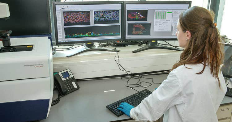
For development and application of tissue models, they use a range of image-based readouts, such as phase-contrast, time lapse, fluorescence microscopy and confocal microscopy, in combination with automated 3D image analysis. The team use the ImageXpress Micro Confocal High-Content Imaging System on a daily basis, serving as their standard high-throughput imager for most in-chip assays. The team has also integrated the ImageXpress Pico Automated Cell Imaging System into their workflow, using it to capture and review images from the entire course of an OrganoPlate® experiment. The user interface of the ImageXpress Pico makes it simple to observe and track each chip for every timepoint and assay captured.
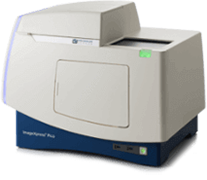
ImageXpress Pico Automated Cell Imaging System
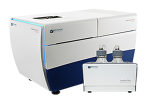
ImageXpress Micro 4 High-Content Imaging System
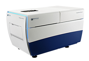
ImageXpress Micro Confocal High-Content Imaging System
Products Used
The ImageXpress® Micro Confocal High-Content Imaging System helps you expand your research with the ultimate combination of speed, sensitivity and flexibility. Capture high quality images of whole organism, thick tissues, 3D spheroid assays, and cellular or intracellular events. Combined with MetaXpress® High-Content Image Acquisition and Analysis Software, the ImageXpress Micro Confocal system provides a complete multi-dimensional, high-throughput screening solution to help you discover your next landmark scientific breakthrough.
The ImageXpress® Pico Automated Cell Imaging System is an affordable, benchtop, cell imager designed for individual biology labs. More than a digital microscope, it combines high-resolution imaging with powerful analysis. Whether running fluorescence imaging or brightfield assays, it features a comprehensive portfolio of preconfigured protocols for cell-based assays to shorten the learning curve, so you can start running experiments quickly.
The Results
In this video, hear from Dr. Bas Trietsch, CTO, MIMETAS, how our ImageXpress Pico and ImageXpress Micro Confocal imaging systems are playing a vital role in the development and analysis of 3D tissue models built on their new OrganoPlate® Graft, an in vitro cell culture microplate platform that allows vascularization of 3D tissues.
References
Wevers, N.R. et al. High-throughput compound evaluation on 3D networks of neurons and glia in a microfluidic platform. Nature Scientific Reports 6, Article number: 38856 (2016).
Junaida A. et al. An End-User Perspective on Organ-on-a-Chip: Assays and Usability Aspects. Current Opinion in Biomedical Engineering. Article in Press (2017).
View our recent webinar with Mimetas: Physiologically-relevant tissue models using a high-throughput Organ-on-a-Chip platform