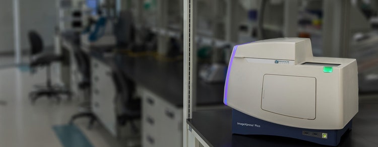
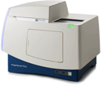
ImageXpress Pico Automated Cell Imaging System
Cellular imaging and analysis system that enables you to go from sample to results in minutes
Digital microscopy with automated brightfield, fluorescence, and Digital Confocal imaging
The ImageXpress® Pico Automated Cell Imaging System is more than a digital microscope, combining high-resolution imaging with powerful analysis. Whether running fluorescence imaging or brightfield assays, the automated imager features a comprehensive portfolio of preconfigured protocols for cell-based assays to shorten the learning curve, so you can start running experiments quickly. With features such as Digital Confocal* 2D on-the-fly deconvolution, Autofocus, Live Preview, multi-wavelength cell scoring, and optional IN Carta® Image Analysis Software workflow, the ImageXpress Pico offers you the ability to advance your discoveries in a small, affordable imager.

Get started quickly
With the icon-driven, user-friendly CellReporterXpress® Image Acquisition and Analysis Software, your entire lab can streamline their digital microscopy. Start capturing and analyzing images with minimal training.

Do more than cell counting
Expand your assays with over 25 preconfigured templates optimized for many cell-based experiments including apoptosis, mitochondrial evaluation, 3D cell models, live cell/timelapse, multiwavelength cell scoring, and neurite tracing.

Automate imaging affordably
Alleviate the hassle of going to the core lab to run your samples. The system’s lab-friendly price allows researchers to afford the convenience of automated imaging and analysis on their lab bench. With options like Digital Confocal, environmental control and z-stack acquisition, the system can be ordered to fit your research.
Features

Multiple imaging modes
The ImageXpress Pico system offers objectives ranging from 4x to 63x and can operate in colorimetric, brightfield, fluorescence, or Digital Confocal 2D on-the-fly deconvolution imaging modes.

Preconfigured analysis protocols
Over 25 preconfigured analysis protocols ranging from simple cell counting to sophisticated neurite tracing analysis. With features like the click-to-find tool, analysis parameters can be optimized by simply clicking on a few cells that match specific criteria.

Plate-to-individual cell view
Data can be visualized at multiple levels from plate overview to individual cells. A wide variety of data visualization tools from the plate and cellular level empower users to learn as much as possible from their images and assays.

Z-stack acquisition
Generate sharper images for more accurate segmentation using z-stack acquisition. Acquire a series of images at different focal points to capture more detail than with a single slice. Users can include all slices or select which slices to include in the final projection.

Quickly and easily identify regions of interest
Live Preview simplifies identification of regions of interest, letting users pan around the sample and interactively adjust focus with a virtual joystick, saving time and effort.

Environmental control
Multi-day, time lapse, and live cell assays can be run using the onboard environmental system with options for humidity, CO2, and O2 control. Optimized to prevent z-drift, the software also provides real-time monitoring of environmental state, ensuring optimal assay conditions.
Experience the powerful combination of the ImageXpress Pico and CellReporterXpress
Latest Resources
Featured Applications
Customer Breakthrough
SUCCESS STORY
Qorium uses the ImageXpress Pico System and SpectraMax iD5 Reader as part of the development of lab-grown leather

