Automated focus algorithms for a variety of applications including custom organ-on-a-chip plates
The ImageXpress Pico system uses two robust autofocus mechanisms:
- Detect Surface – hardware autofocus
- Find Best Plane – image-based autofocus
Hardware autofocus uses an LED beam to find reflective surfaces and is designed for speed. It works well for adherent samples in plates or chamber slides. When enabled, image-based autofocus searches a range for the best focus plane based on image contrast. It works well for slides with a coverslip or for samples in a plate that are not flat, such as suspension cells or spheroids. The ImageXpress Pico system with CellReporterXpress software provides reliable focusing across all of your labware to ensure high-quality imaging for a wide variety of sample types.
Detect surface algorithms
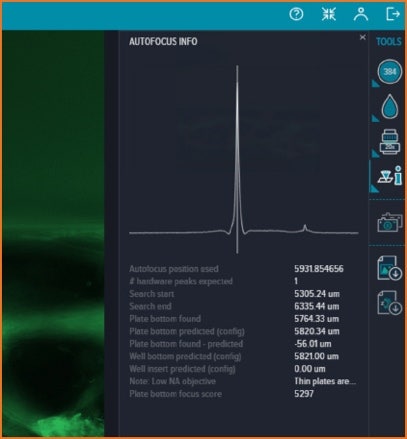
Plate Bottom
Hardware autofocus detects the surface closest to the objective—that is, the plate bottom. It works well for adherent samples in plates or chamber slides when imaging at low magnification or if your labware has a thick bottom. This is the fastest hardware autofocus option.
Well Bottom
Hardware autofocus detects the two surfaces closest to the objective—that is, the plate bottom and well bottom. The Well Bottom option is designed for samples in a liquid medium, such as well plates or chamber slides. This is the most commonly used hardware autofocus option.
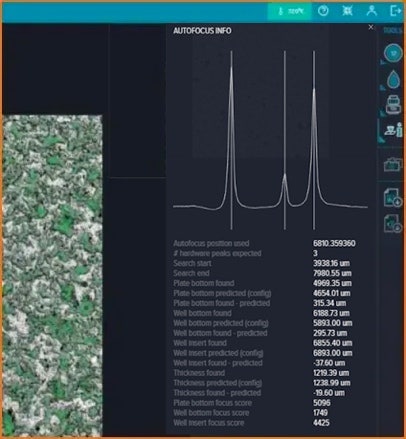
Well Insert
Hardware autofocus detects the three surfaces closest to the objective—that is, the plate bottom, well bottom, and well insert. The Well Insert option is designed for well inserts in well plates or any labware design that has a distinct third surface.
Find best plane algorithms
If the Detect Surface options do not provide satisfactory focus, select the Find Best Plane option to add image-based autofocus for the first channel within the selected search range. Adding image-based autofocus is useful for thicker samples, samples with variable best focus planes, or labware with variable thickness.
The Find Best Plane algorithms center on the surface detected by the hardware autofocus and search within a specific range to find the optimal focal plane.
- Normal Search Range – searches a range of 7.5% above and 7.5% below the labware bottom
- Wide Search Range – searches a range of 20% above and 20% below the labware bottom
- Superwide Search Range – Unlike these two options, which search around the surface, the Superwide Search Range searches a range of 300 μm above that surface.
Anchor focus position
For fastest screening speed, we have introduced the ability to select the Anchor Focus Position to save current focus position and disable the autofocus controls. The software uses the saved focus position for any ensuing snaps of preview images and for acquisition. This can dramatically speed the acquisition of plates at low magnification or when you are working with macroscopic samples like whole organisms or tissues.
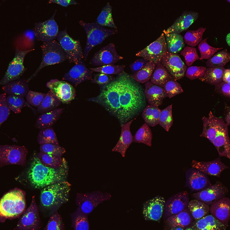
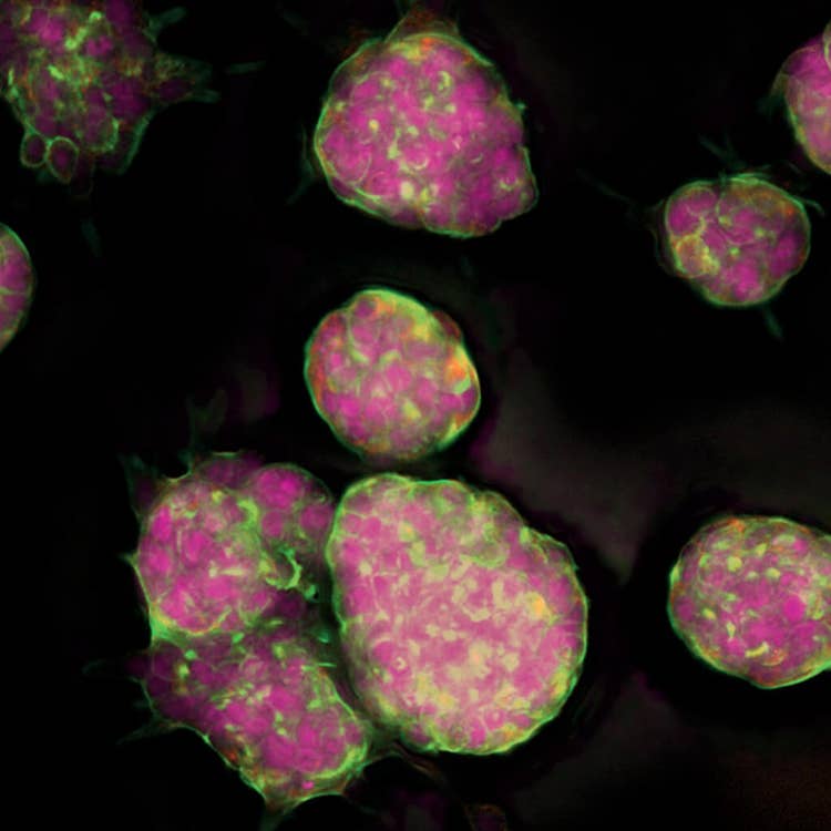
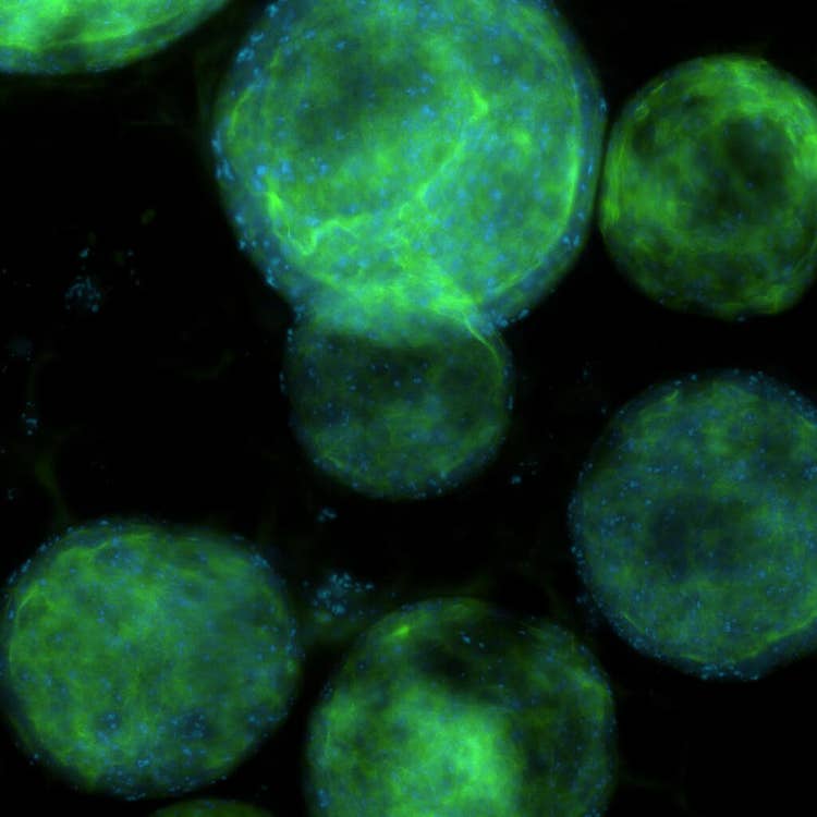
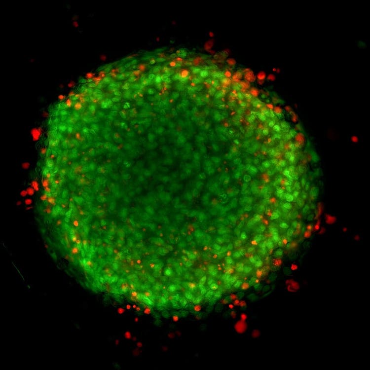
Cellular image gallery


†Data and images were acquired during development using customer samples. Results may vary. Highlighted features’ price, time to deliver, and specifications will vary based on mutually agreed technical requirements. Solution requirements may cause adjustment to standard performance.
Latest Resources
Resources of Autofocus
Blog
Innovation at Molecular Devices: Updates in Automated, High-Content Imaging
From customer feedback to workflow improvements The path to understanding complex biological processes and diseases is paved with a lot of challenges. As the desired level of…
Brochure
CellReporterXpress Image Acquisition and Analysis Software
The CellReporterXpress® Automated Image Acquisition and Analysis Software works with the ImageXpress® Pico Automated Cell Imaging System. It has a clean, easy-to-learn interface for per…
eBook
Transform your cell imaging
From endpoint and live-cell imaging applications to imaging and analyzing 2D monolayers, 3D cell models, and organ-on-a-chip platforms, the world of cellular imaging is very diverse. Th…
Blog
Tips for running a successful live cell imaging experiment
There have been significant advancements in microscopy and camera technology, as well as advancements in technologies for labeling molecules of interest over the past decade. These…
Publications
Disease Modeling with 3D Cell-Based Assays Using a Novel Flowchip System and High-Content Imaging
There is an increasing interest in using three-dimensional (3D) cell structures for modeling tumors, organs, and tissue to accelerate translational research. We describe here a novel au…
Application Note
Quantification of spheroid growth and morphology
There is a growing trend to develop screening assays using three-dimensional (3D) cell culture.
Publications
3D Microscopy Keeps Getting Faster, Smarter, Leaner
With each passing day, continuous advancements are being made in the studies that cutting-edge instruments can perform and in the complexity of the biological samples that are being exa…
Publications
From 3D high throughput to simple automated imaging: Cellular imaging within reach of every laboratory
OUR ABILITY to image cells has come a long way since the pioneering days of Galileo Galilei and Antonie van Leeuwenhoek. The multiple imaging techniques available today range from simpl…
Publications
Everything you need to know about the ImageXpress Pico Automated Cell Imaging System
Depending on your application and exactly what you are researching, there are multiple imaging techniques available, ranging from confocal microscopy to electron microscopy and more - e…
Application Note
Monitor multiple stages of apoptosis with live cell kinetic imaging
The study of apoptosis is a critical aspect of drug discovery and development. Additionally, studying the relationship between apoptosis and other factors, such as oxidative stress, is…