
Cell Painting
A high-content, image-based assay for morphological profiling using multiplexed fluorescent dyes
What is Cell Painting?
Cell Painting is a high-content, multiplexed image-based assay used for cytological profiling. In a Cell Painting assay, up to six fluorescent dyes are used to label different components of the cell including the nucleus, endoplasmic reticulum, mitochondria, cytoskeleton, Golgi apparatus, and RNA. The goal is to “paint” as much of the cell as possible to capture a representative image of the whole cell.
Automated image analysis software is used to extract feature measurements from each cell. The number of unique measurements is usually in the range of 100 to 1000 per cell. These measurements typically include intensity, texture, shape, size as well as the proximity of an object to its neighboring structure, which provides an indication of the spatial relationship between organelles. Together, these measurements form the phenotypic profile.
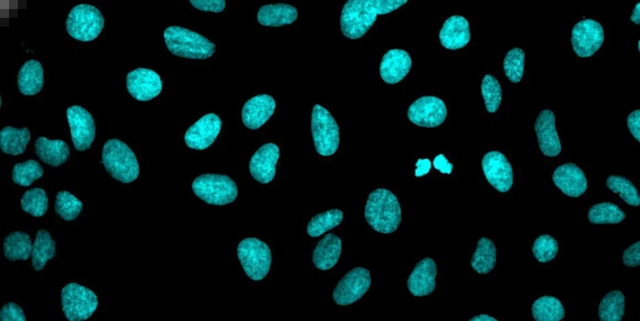
Nucleus
Dye: Hoechst 33342
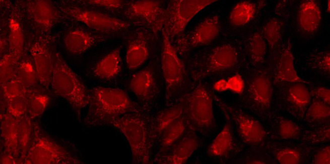
Mitochondria
Dye: MitoTracker Deep Red
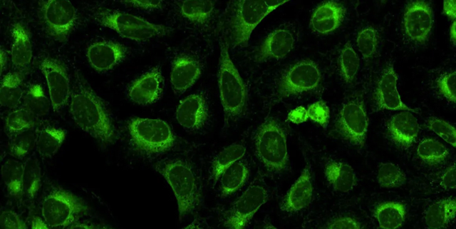
Endoplasmic reticulum
Dye: Concanavalin A/Alexa Fluor 488 conjugate
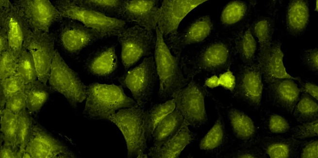
Nucleoli, cytoplasmic RNA
Dye: SYT0 14 green fluorescent nucleic acid stain
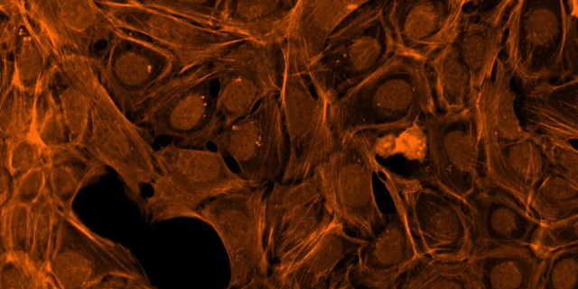
F-actin cytoskeleton, Golgi, plasma membrane
Dye: Phalloidin/Alexa Fluor 568 conjugate, wheat-germ agglutinin/Alexa Fluor 555 conjugate

Composite image consisting of Red: Actin and golgi; Green: ER; and Blue: nuclei
Cell Painting for phenotypic profiling
The phenotypic profile of a cell reveals the specific biological state of a cell. More specifically, it can be used to interrogate biological perturbations because the cellular morphology is influenced by factors such as metabolism, genetic and epigenetic state of the cell, and environmental cues. In addition, it can be used to characterize healthy cells from diseased cells. Because a phenotypic profile is an aggregation of a large number of measurements, it is more sensitive to deviations or changes to those features extracted from Cell Painting assay. In other words, a phenotypic profile can capture certain characteristics of the cells that may not be obvious to the naked eye.
In an interview with the Science Explorer, Angeline Lim, PhD., explained, “I like to think of Cell Painting or phenotypic profiling as analogous to facial recognition. It's a little bit like how Facebook or iPhoto tags people. Once you have a photograph and you tag it, the software in the background extracts information about the photo and calculates a profile. When another photo pops up, the software compares its profile to that of the previous photo to see if they are the same or different. This is basically how Cell Painting works. With Cell Painting, we expect the image analysis part to cluster like cells from unlike cells, or diseased cells from healthy cells. This potentially has many applications, especially in drug discovery.”
Examples of treated cells and phenotypic changes
Here is an example in which treated cells show obvious changes in their phenotypes—only three of the five wavelengths are shown in this composite: nuclei in blue, endoplasmic reticulum in green, actin & Golgi in red.
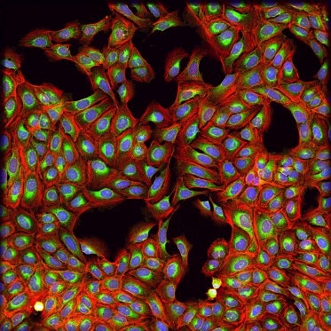
Control, untreated cells.
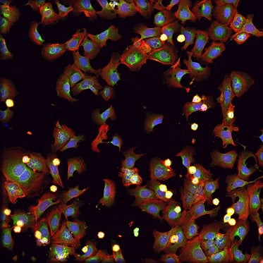
Cells treated with rotenone, a toxin frequently used in insecticides. It is also known to inhibit mitochondrial ATP production and has been shown to have anticancer activity in various cancer cells.
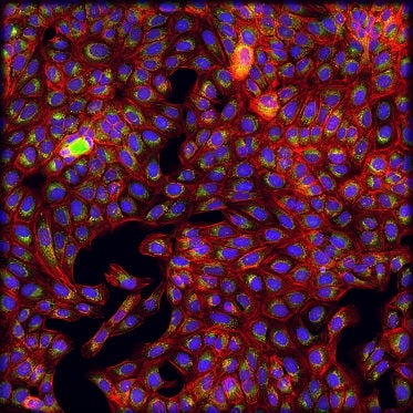
Cells treated with chloroquine. Chloroquine was first developed for malaria treatment.
General workflow for Cell Painting assay
One of the advantages in the Cell Painting assay is that the overall workflow is one that is familiar to many biologists. First, you plate the cells. Next, you introduce a type of perturbation; it can be chemical or genetic (e.g., adding of compounds, small molecules, or an RNAi library). After a suitable incubation period, the cells are stained with a set of Cell Painting dyes. It is also possible to use other dye combinations that are more suited to your specific assay.
Once the cells are “painted,” cell images are acquired with a high-content imager such as our ImageXpress® Confocal HT.ai, the latest addition to our portfolio of high-content imaging system. An automated image analysis software like our MetaXpress® or our IN Carta image analysis software is used for feature extraction where cells and their components are identified and measured. Finally, the measurements are further processed using various data analysis tools to create and compare phenotypic profiles, perform clustering analysis, and identify targets to derive the morphological profiles.
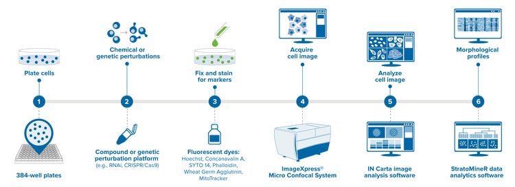
- Plate cells into labware (384-well plate)
- Treat cells with chemical or genetic perturbation (e.g., RNAi, CRISPR,/Cas9) or viruses.
- Stain with fluorescent dyes (e.g., Hoechst, Phalloidin, MitoTracker)
- Acquire cell images with high-content imaging system
- Analyze cell images to extract features and measurements using automated image analysis software
- Derive morphological profiles from measurements
Learn more about Cell Painting
Cell Painting is emerging as a valuable tool with its many important potential applications particularly in drug discovery. Learn how to optimize high-content imaging capabilities and perform multiplexed assays using preconfigured or custom modules from MetaXpress software to provide a data rich, phenotypic profile.