QuickID Targeted Image Acquisition
QuickID Targeted Acquisition
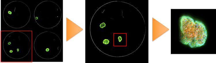
QuickID™ Targeted Acquisition works by acquiring an entire field-of-view (FoV) of a sample at low magnification, finds objects of interest or rare events through a fast image analysis process, and then re-acquires only these wells or sites at high magnification with a user configured protocol that may include the relevant wavelengths, z-stacks, and time-points.
QuickID Targeted Acquisition is now integrated into the MetaXpress Acquire software, providing a simple and intuitive workflow. Regions of interest can be identified using the AI-powered analysis tools form IN Carta Image Analysis software to enable targeted imaging in the most challenging of assays.

Increase imaging efficiency using QuickID Targeted Acquisition
Investigators performing image-based high content screening may face several technical challenges if they need to image their samples at high resolution, at multiple wavelengths, and over a range of z-planes. Such experiments often have lengthy acquisition times (between several hours and a couple of days), a vast need of image storage space (100s of GBs), and high computational power requirements to keep image analysis time managable (the fewer hours the better).
A simple way to minimize all of these challenges is to acquire only the fields-of-view on the slide or in the microplate that contain objects of interest. This technique can be especially useful when the object of interest is scarcely abundant in each sample, randomly distributed in space, or only occurs as a rare event in some of the microwells. This solution, when put into use with our high-content imaging systems and image analysis software, dramatically reduces the time-to-result as well as image storage requirements.
Acquire data 10X faster from 3D cell cultures
In the application note “Acquire data 10X faster from 3D spheroids cultured using Symphony® and VersaGel®” a light crosslinkable hydrogel was used as a matrix to grow 1-4 spheroids/well in a 3 dimensional space. After staining, QuickID was performed using a 2X objective to capture the entire well in one field-of view. Objects of interest were identified and their X and Y coordinates automatically referenced so the ImageXpress Micro system obtained only relevant images at 10X magnification in three wavelengths across multiple z planes. With QuickID, acquisition time was 10X faster than imaging all sites in every well at high magnification and this also reduces the required storage space by 20X.
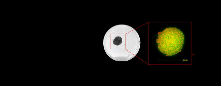
QuickID was used to streamline spheroid image acquisition. An image acquired at low magnification to view the entire well in one field-of-view was used to identify objects for automatic re-imaging at higher magnification using three wavelengths across multiple z planes.
Automatically image 3D spheroid in the center of FoV
There are several methods for culturing spheroids for screening antitumor drugs. In some formats, the spheroids are not always centered in the well, which may make it difficult to acquire the object of interest in a single field-of-view (FOV) at high magnification. QuickID allowed us to automatically image each spheroid in the center of the FOV using a 20X water immersion objective to collect z stacks in multiple wavelengths without acquiring extra sites.

The images were acquired on the ImageXpress Micro system. QuickID was performed with a 10X objective to quickly image an entire microplate. Spherical objects were then identified and their X,Y coordinates were used to automatically acquire only sites that contained a tumor microtissue with a 20X water immersion objective in multiple wavelengths and z planes. Image analysis was performed to find and measure objects within the 3D volume.
Other applications for targeted acquisition
QuickID streamlines the process so only relevant images are acquired which results in significant time-savings and reduced storage needs. Examples of other application areas that would benefit from the use of QuickID include live imaging of dividing cells or model organisms, and imaging specific areas of tissue sections on a slide.