
Cell counting using automated cell imaging
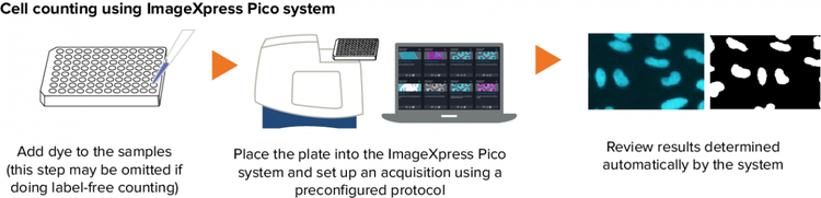
Cell counting is fundamental and critical to numerous biological experiments. Assays such as drug compound toxicity, cell proliferation, and inhibition of cell division have a need to assess the number or density of cells in a well. Automated imaging can greatly speed up the cell counting process while reducing manual labor and human errors. For example, users can simply place the sample into the ImageXpress Pico Automated Cell Imaging System, and after a simple setup in the software, the system can count cells automatically, allowing users to have walkaway time. Cells can be counted using a variety of methods, such as label-free cell counting under transmitted light or detection of nuclear dye with fluorescent imaging.
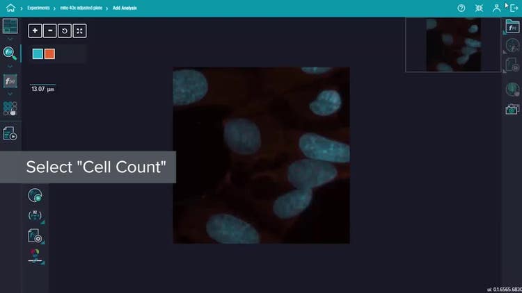
Analyzing cell counting is quite easy in the ImageXpress Pico system. Simply select the "Cell Count" protocol, and the software will calculate the value automatically. If you are using a well plate, the analysis is automatically run across the plate with no extra steps. To visualize results, the user can turn on and off the mask.
Lastly, in addition to cell count, automated imaging also provides data-rich outputs such as nuclear size, total and average cell area, and intensity. With the ImageXpress Pico system, data can be presented in numerous formats including image montages, heat maps, scatter plots, bar graphs, and more. These data are also easily exportable for downstream analysis or presentations.
In summary, the benefits of using automated imaging for cell counting include:
- Reduce manual labor, thus allowing walkaway time
- Minimize human error
- Choose between label-free cell counting or fluorescent cell counting
- Obtain data-rich outputs such as nuclear size, total and average cell area, and intensity