
Apoptosis analysis using automated cell imaging
Apoptosis is a process of programmed cell death important to a number of biological processes including embryonic development and normal tissue maintenance. Disruption in the regulation of apoptosis has been implicated in various diseases including cancer. Biochemical events lead to characteristic changes in cell morphology such as cell shrinkage, nuclear fragmentation, chromatin condensation, and mRNA decay, and ultimately cell death. Apoptosis can be initiated via the intrinsic or extrinsic pathways, and both pathways induce cell death by activating caspase enzymes.
One way to analyze apoptosis is to detect caspase-3 and caspase-7, both which are activated during the execution phase of apoptosis. For example, the EarlyTox™ Caspase-3/7 NucView 488 Assay Kit offers a substrate that can detect caspase-3/7 activity within intact cells. The substrate consists of a fluorogenic DNA dye that is coupled to the caspase-3/7 DEVD recognition sequence. Initially nonfluorescent, the substrate can permeate the plasma membrane and enter the cytoplasm. In apoptotic cells, caspase-3/7 cleaves the substrate, releasing a high-affinity DNA dye that migrates to the cell nucleus and binds to DNA. Excitation at 500 nm results in an emission of a bright green fluorescence at 530 nm and can be used to evaluate the level of caspase activation. Apoptotic cells can then be visualized and quantified using automated imaging.
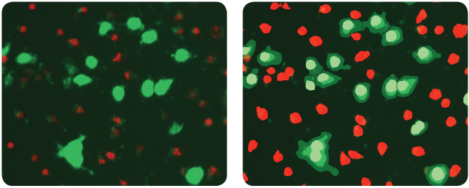
HeLa cells were treated with the compound staurosporine and stained with EarlyTox Caspase-3/7 NucView 488 Assay Kit (green). The overlay mask (right) shows apoptotic cells identified in green and non-apoptotic cells in red.
Analysis of apoptotic cell images can be performed on the ImageXpress Pico Automated Cell Imaging System, which offers a preconfigured "Apoptosis" protocol template designed for easy image acquisition and analysis. Useful output such as number and percentage of apoptotic cells and live cells can be calculated automatically. Below is a typical workflow for imaging and analyzing apoptosis assay plates.
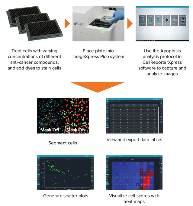
A typical workflow for imaging and analyzing apoptotic cells.
In summary, the benefits of using the ImageXpress Pico system for apoptotic assays include:
- Allow direct visualization and monitoring of cell health
- Enable objective assessment of cells, thus leading to reproducible results
- Determine valuable outputs such as number and % of apoptotic cells automatically
To learn more, download our application note "Analysis of apoptosis using automated cell imaging" >>