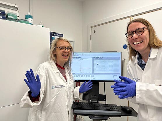
Dementia Research Institute at Cardiff University uses the FLIPR Penta System to study the role of microglia in Alzheimer’s disease

“The FLIPR Penta surpasses the efficiency of an inverted microscope by enabling me to image a significantly larger quantity of cells and samples. Remarkably, it allows me to capture approximately 100 times more samples daily.”
Dr. Emily Maguire
COMPANY/UNIVERSITY
Cardiff University, Dementia Research Institute
TEAM MEMBERS
Dr. Emily Maguire,
Dr. Nina Stöberl,
Dr. Hazel Hall-Roberts,
Dr. Bethany Shaw,
Ms. Rachel O’Donoghue,
Ms. Jincy Winston
PRODUCTS USED
FLIPR Penta High-Throughput Cellular Screening System
384-well head for FLIPR Penta
FLIPR Calcium 6 Assay Kit
The Challenge
Dr. Emily Maguire and her team at the UK Dementia Research Institute at Cardiff University aim to identify the causes of Alzheimer's disease that will improve the care, diagnosis, and, ultimately, treatment of patients.
Research in Emily’s group focuses on identifying what’s different between the brain cells of people with and without Alzheimer’s disease. Specifically, they are examining a distinctive class of brain immune cells known as microglia, which have emerged as crucial contributors to the progression of Alzheimer's disease. They use genetic testing to estimate Alzheimer's risk and collect blood samples from two groups: low-risk individuals and high-risk individuals already affected by the disease. The blood cells are transformed into stem cells and then differentiated into microglia. This approach enables the team to compare microglia from high-risk and low-risk individuals and identify differences related to Alzheimer’s disease. This research would allow scientists to develop specific therapies for Alzheimer’s disease in the future.
“To image one sample on the inverted microscope with the same number of repeats as on the FLIPR Penta, it would take me around 3 hours of hands-on time (plus 1.5 hours for calcium dye loading). However, with the FLIPR Penta, you can image 30x more samples in approximately 30 minutes of hands-on time (plus calcium dye loading). This significantly simplifies the process of obtaining repeats.”
– Dr. Emily Maguire
The Solution
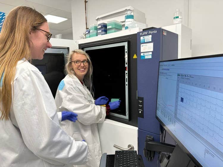
The team utilized the FLIPR Penta to compare the response of microglia from patients with high Alzheimer's risk to those from patients with low Alzheimer's risk when exposed to known calcium modulators. These comparisons aimed to identify discrepancies in calcium signaling between diseased and non-diseased cells, potentially shedding light on the underlying pathology.
The approach involved plating cells in a 384-well plate and staining them with a green fluorescent Calcium 6 dye from Molecular Devices. After incubation, they loaded a specific compound (e.g., ATP) into another plate. Both plates were placed in the FLIPR Penta, which added the compound to the cells and recorded changes in fluorescence both before and for 5 minutes following the addition of the compound. This enabled the team to measure the calcium released upon application of the compound and, thus, the level of cell signaling. To standardize the results, the team also captured an image of the cell plate and normalized the fluorescence measurements based on the cell number in each well, enabling them to obtain accurate and reliable comparisons between wells.
“The FLIPR Penta allows us to assay a large number of samples, up to 24 per experiment in a 384-well plate, while minimizing variability resulting from fluctuations in external factors between experiments. Moreover, the FLIPR instrument allows us to administer precise quantities of reagents (specifically ATP in our case) to each well simultaneously, thus ensuring uniformity across all wells and enhancing consistency in our measurements.”
– Dr. Emily Maguire
It takes the researcher roughly 2 hours to prepare a plate for a FLIPR calcium assay, although much of this time is just dye incubation. The actual imaging of a plate takes 10 minutes. For this reason, by staggering staining with Calcium 6, it would be possible to image over 50 plates a day.


Products Used
FLIPR Penta High-Throughput Cellular Screening System and FLIPR Calcium Assay Kits
The Results
Whilst the research is not conclusive, preliminary results showed a potential disparity in ATP-mediated calcium signaling between the two groups.
“By conducting further experiments using the FLIPR Penta technique with an expanded range of patient-derived cell lines, we can enhance the robustness of our study and potentially obtain more conclusive results.”
– Dr. Emily Maguire
If significant variations are observed, it would suggest that ATP-calcium signaling plays an important role in Alzheimer's disease, potentially opening up new avenues for the development of therapies.

A. The graph shows how there was a non-significant trend towards increased calcium release following the addition of ATP in our high Alzheimer’s risk microglia in comparison to the low-risk cells. This data was collected with a preliminary set of 9 stem cell lines (6 high risk, 3 low risk). The different colors indicate cells from various individuals, and the different shapes indicate different experimental replicates.
B. Fluorescent microscopy image of microglia stained with the Calcium 6 dye.
Besides ATP, the team identified several other mediators within cells that play a crucial role in calcium mobilization. Hence, in future investigations, they can explore the effects of introducing different molecules known to induce calcium release through various mechanisms, such as other nucleotides like ADP and UDP that bind to different receptors. The idea is that by comparing the responses of microglia with high and low risk of Alzheimer's disease to these alternative activators, the team can assess if any distinctions exist between the two groups.
Find out more about the UK Dementia Research Institute at Cardiff University on their website
Assaying Microglia Functions In Vitro | UK Dementia Research Institute