GPCRs (G protein-coupled receptors)

GPCRs (G protein-coupled receptors)

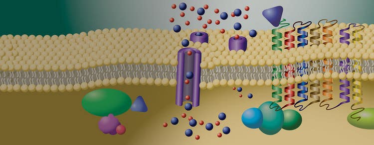
GPCRs or G protein-coupled receptors are proteins located on the cell surface that recognize extracellular substances and transmit signals across the cell membrane. GPCRs do this by activating guanine nucleotide-binding proteins (G protein) that are responsible for signal transduction inside the cell. The signal delivery is very important in various cellular responses including cell growth, gene transcription, post-translational changes, and communication with other cells. This brings on adjustments in the body to adapt to the environmental changes such as increased heart rate when feeling threatened or change in vision in response to dim light.

The human genome alone consists of at least 1,000 different GPCRs that detect hormones, lipids, amines, neurotransmitters, and light, among other things.

A GPCR consists of three regions. The extracellular part detects and binds the ligand. Then, the seven-transmembrane region undergoes a conformational change. Finally, this change activates the C-terminus that triggers the corresponding G-protein.


cardiomyocytes screening

iPS cell-derived cardiomyocytes are especially attractive cell models because they represent gene expression profiles as well as phenotypic characteristics similar to native cardiac cells.

G protein-coupled receptors and ion channels


GPCRs are the largest protein family, with between 600 and 1000 members, and have been linked to many normal biological as well as pathological conditions. They are also known as seven transmembrane (7-TM) receptors, and about 45% of modern medicinal drugs affect this target class. The function of GPCRs is highly diverse, recognizing a wide range of ligands, including photons, small molecules, and proteins.

Ion channels are pores in the cellular membrane that allow ions to pass in and out of the cell. There are over 400 genes for ion channels in the human genome. Many of them have been targeted by drugs that are now blockbusters. Direct measurement of ion channel activity is measured using traditional electrophysiology equipment for patch-clamping. However, the throughput is very low. Ion channel activity can also be measured indirectly with much higher throughput by using fluophores sensitive to changes in membrane potential, calcium flux, and potassium flux.


Monitor GPCR activity for drug discovery


Changes in GPCR activity lead to abnormalities in cellular signaling pathways, which result in inflammation, cardiovascular diseases, mental disorders, hormonal imbalances, and cancer. That’s why GPCRs are at the center of drug discovery, with approximately 34% of all FDA-approved drugs targeting 108 well-defined GPCRs.

Various assays can be used in drug discovery to monitor GPCR activity and corresponding intracellular changes.

Calcium is an important messenger triggered by GPCR activity. Therefore, changes in intracellular calcium signaling are strong indicators of the activation state of GPCRs. Calcium flux assays can be used to monitor intracellular calcium levels in drug screening.

Monitoring calcium oscillations is also crucial to predicting in vitro toxicity of your drug candidates.

Cyclic Adenosine Monophosphate (cAMP) is another important messenger involved in signal transduction pathways. The change in intracellular cAMP levels indicates the specific GPCR-G Protein coupling. cAMP assays can give valuable information about the GPCR subtypes.

It is also possible to monitor the GPCR activity through transfluor assays, where the main focus is on GPCR desensitization upon ligand binding. These assays can be implemented in drug screening to monitor GPCR activation/deactivation and movement across the cell membrane.


Solutions for identifying early leads against GPCRs


We offer a variety of assay and instrument solutions to support studies of GPCR and ion channel function including assay kits, cellular screening and imaging systems, and microplate readers.

Here we focus on applications using the FLIPR High-Throughput Cellular Screening System, the Screenworks Peak Pro 2 software module along with various FLIPR assay kits to provide a high-throughput kinetic screening solution for toxicology and lead compound identification.
 

  • Cardiomyocyte

    Cardiomyocyte

    Early prediction of drug-induced functional cardiotoxicity requires robust in vitro systems suitable for high-throughput screening. Readily-available iPSC-derived human cardiomyocytes may be used in conjunction with calcium-sensitive dyes, with beat rates and patterns monitored aschanges in intracellular calcium in response to both GPCRs and ion channels. These calcium peaks can be analyzed with our ScreenWorks Peak Pro 2 software which provides the investigator a suite of powerful tools for quantification of cardiomyocyte activity.

    Read application notes:

    Cardiotoxicity: calcium oscillations

    Cardiotoxicity: Calcium oscillations

    Induced pluripotent stem cell (iPSC)- derived cardiomyocytes are a particularly attractive in vitro model system due to their use for evaluation of compound effects on both cardiac function and safety. Calcium signal oscillation reflects changes in cytoplasmic calcium concentration making it possible to use a calcium sensitive dye such as the EarlyTox Cardiotoxicity Kit.

    Read application notes:

  • Evaluation of calcium flux

    Calcium Flux

    The evaluation of calcium flux is a long accepted, tried and true measure of cellular activity. Calcium flux can be used as a measurement for a host of cellular processes including neurotransmitter release, GPCR activity, ligand gated ion channels, and cardiomyocyte beat patterns, among many others. The FLIPR System is the drug discovery tool for evaluation of calcium flux in high and ultra-high throughput screening due to its ease of use, sensitivity and user configurability.

    Learn more:

    Fura-2 QBT calcium

    Fura-2 QBT Calcium

    Fura-2 dye has long been considered an important tool to measure calcium mobilization in cellular imaging, GPCR mediated intracellular calcium flux, and ion channel activation. This ratiometric dye helps correct for assay inconsistencies in dye loading or cell plating through calculating the fluorescence intensity ratio between bound and free indicators. However washing is required, increasing well-to-well variability and adding time and complexity to each assay.

    The Fura-2 QBT™ Calcium Kit from Molecular Devices incorporates proven quench based technology with a ratiometric Fura-2 calcium indicator to provide a homogenous assay to minimize cell based variability, while increasing throughput through elimination of cell washing prior to detection.

    Learn more:

  • Homogeneous solution for GPCR assays

    Homogeneous solution for GPCR assays

    Cell-based assays have become an indispensable method for screening and compound profiling in the early drug discovery process. To date, such assays have proven to be some of the most reliable and reproducible methods in receptor characterization studies, primary screening campaigns and compound profiling programs. For Gq-coupled GPCR targets specifically, homogeneous fluorescent calcium flux assays with masking technology are the methodology of choice.

    Combining a novel fluorophore and proven masking technology, the FLIPR Calcium 5 Assay Kit delivers reliable pharmacology, a larger signal window, and improved assay performance. With the FLIPR Calcium 5 Assay Kit and the FLIPR System, consistent screening of a variety of receptors and targets, especially those with small calcium signal responses, can be obtained in an easy-to-use, homogeneous format.

    Read Application Note:

    Live cell Gi - and Gs-coupled GPCR second messenger signaling

    GPCR second messenger signaling

    Detection of Gi and Gs -coupled GPCR second messenger signal activity has been traditionally accomplished using assays such as radioactive binding or endpoint cAMP assays that require cell lysis. Such assays measure activity at a single time point in the cellular response and do not provide kinetic information. Another option utilizes forced-coupling of Gi and G-coupled GPCRs to Gα16 followed by fluorescence detection of calcium flux upon agonist receptor activation. Again, this assay is sub-optimal as it does not signal through the biorelevant cAMP pathway.

    Here we demonstrate endogenous receptor activity in CHO-K1 and HEK-293 cell lines stably expressing the GloSensor plasmid using the FLIPR System.

    Read Application Note:

  • Live cell kinetic assays

    Live Cell Kinetic Assays

    The ImageXpress Confocal and Micro 4 High-Content Imaging Systems offer optional fluidics and environmental control modules, enabling single-channel pipetting for compound addition and media exchange during rapid kinetics or extended time-lapse experiments. The Transfluor Cell-Based GPCR Assay Kit is a valuable tool used during high content screening (HCS) to track G-protein coupled receptor (GPCR) activation by quantifying GPCR desensitization and recycling using GFP-labeled ß-arrestin with automated image analysis.

    Read Application Note:

    Measure intracellular calcium flux

    Comparison of intracellular calcium measurements

    G protein-coupled receptors (GPCRs) play an important role in cell signaling. When the receptor is activated by a ligand, receptor conformation is changed, triggering G-protein activation inside the cell. An active G protein has the potential to induce various cascades of intracellular messengers including calcium. The FLIPR System performs high-throughput, functional cell-based assays and is the system of choice in drug discovery for evaluating changes in intracellular calcium detected through use of fluorescent calcium-sensitive reporter dyes. Here we provide a basic protocol for performing a calcium mobilization assay on the FLIPR System using the FLIPR® Calcium Assay Kit, a homogeneous, fast and reliable fluorescence assay.

    Read Application Note:

  • Monitoring Gq-coupled protein receptor activity

    Monitoring Gq-Coupled Protein Receptor Activity

    Gq protein-coupled receptor activation is commonly monitored in live cells in real time using calcium-sensitive dyes on a fluorescence plate reader. Automated liquid handling within the plate reader is generally required to deliver agonist compounds to the cells in the microplate while the detection system takes real-time readings of compound-induced changes in fluorescence intensity values. Analysis of the resulting kinetic readings yields information about the compound response profiles, including EC50 and IC50 values for agonists and antagonists.

    Read Application Note:

    Neuronal assays

    Neuronal Assays

    The FLIPR System is an excellent tool for monitoring calcium flux related to neuronal activity. Newly developed commercially available iPSC neurons have made high-throughput neuronal screening a reality. These cells and assays are essential for monitoring a variety of factors including neural cell culture conditions, drug compound effects, and the effect of environmental neurotoxins. Neuronal activity and neurotransmitter release can be evaluated with fluorescence-based calcium indicator dyes combining fast, simple, and powerful assays with scalable throughput.

    Learn more:

Latest Resources

Resources of GPCRs (G protein-coupled receptors)

Related Products & Services of GPCRs (G protein-coupled receptors)