ImageXpress Nano Automated Imaging System [Discontinued]
This product has been discontinued.
The ImageXpress® Nano Automated Imaging System is no longer available for purchase. Support for existing customers may continue per applicable service agreements.
For service, documentation, or installed‑base support, please visit SpectraNet.
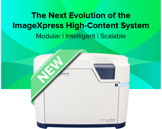
Introducing ImageXpress HCS.ai High-Content Screening System — Modular, intelligent, and built for the future of high-content imaging
Building on the trusted legacy of the ImageXpress Nano system, Molecular Devices introduces the next evolution in high-content imaging — the ImageXpress HCS.ai High-Content Screening System. This fifth-generation modular platform is designed to scale effortlessly with your research, empowering you to move seamlessly from widefield imaging to advanced laser-based confocal modalities—all within one adaptable system.
Engineered for discovery without limits, ImageXpress HCS.ai system combines the renowned optical excellence you trust with revolutionary new capabilities: accelerated acquisition speeds, the intuitive, redesigned MetaXpress® Acquire software, and next-generation, AI-driven IN Carta® analysis to extract deeper insights from complex 2D and 3D models.
Whether you're progressing from widefield microscopy to confocal or expanding into advanced, AI-powered 3D analysis, the ImageXpress HCS.ai system delivers the speed, flexibility, and intelligence to keep your research at the cutting edge—built upon the innovation you already trust.
ImageXpress Nano: Fluorescence imaging platform within reach of every lab
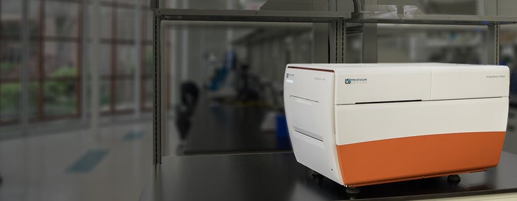
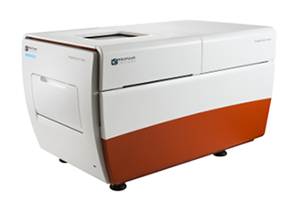
The ImageXpress® Nano Automated Imaging System features a long life, solid state, light engine, and optics to reliably deliver the right assay sensitivity. Capture fine details of a variety of cellular and subcellular assays with this powerful and flexible fluorescent microscopy solution. The system includes MetaXpress® High-Content Image Acquisition and Analysis Software with tools for 2D and 3D imaging and time lapse analysis, as well as a range of needs from ease-of-use through to proprietary assay design.

Image label-free
Brightfield imaging allows for rapid acquisition without the use of harmful fluorescent agents.

Streamline image analysis
The modular toolbox in the MetaXpress® software allows for the quick setup of hundreds of routine assays. Choose from our optional selection of turnkey application modules for greater convenience.

Capture a diverse range of samples
With 2x to 60x magnification, the system offers the flexibility to image whole-well (C. elegans, zebrafish), as well as sub-cellular details (vesicles, organelles).
Features

Large field of view
An entire well of a 384-well plate can be captured in a single image at 4x magnification for faster throughput.

Automated Stages
Fully automated X, Y, and Z stages with resolution better than 25 nm.

Wide range of filters and objectives
The system can be configured with different filters or objectives (2-60x) to meet research needs.

Five fluorescent channels
The system can have up to 5 fluorescent filters installed at one time. The software allows up to 7 channels to be acquired at one time which enables multi-channel fluorescent and transmitted light imaging in one experiment.
High-speed autofocus
Laser autofocus enables quick, consistent focusing across plates, slides and uneven surfaces.
Environmental control option
Multi-day, time lapse and live cell assays can be run using the onboard environmental system with options for temprature, humidity, and CO2 control.
