Yashraj Biotechnology leverages the ImageXpress Nano to substantially improve their multi-parametric imaging and analyses
COMPANY/UNIVERSITY
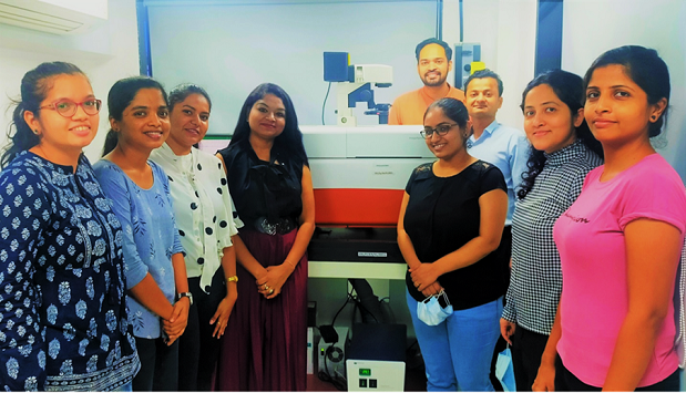
Yashraj Biotechnology Limited
TEAM MEMBERS
Dr. Shweta Bhatt, Chief Scientific Officer and R&D Head
PRODUCTS USED
ImageXpress® Nano Automated Imaging System
SpectraMax® iD5 Multi-Mode Microplate Reader
MetaXpress® High-Content Image Acquisition and Analysis Software
The Challenge
Unexpected toxicity in areas such as cardiotoxicity, hepatotoxicity, and neurotoxicity is a serious complication of clinical therapy and one of the key causes for failure of promising drug candidates in development. Animal studies have been widely used for toxicology research to provide preclinical security evaluation of various therapeutic agents under development. Species differences in drug penetration of the blood-brain barrier, drug metabolism, and related toxicity contribute to failure of drug trials from animal models to human. The existing system for drug discovery has relied on immortalized cell lines, animal models of human disease, and clinical trials in humans. Moreover, drug candidates that are passed as being safe in the preclinical stage often show toxic effects during the clinical stage. Only about 16% of drugs, on average, are approved for human use.
Before switching to the ImageXpress Nano, Yashraj Biotechnology used a phase contrast and fluorescence microscope, but found they lacked the ability to take z-stacks of spheroids/organoid models and didn’t have any image analysis software.

- Dr. Shweta Bhatt, Chief Scientific Officer, Yashraj Biotechnology
The solution
Yashraj Biotechnology is developing cutting-edge research in the stem cell domain and the ImageXpress Nano Automated Imaging System has enabled them to better image their in-house-developed iPSC-derived hepatocytes, cardiomyocytes, and neural cells, as well as several different types of multiparametric assays.

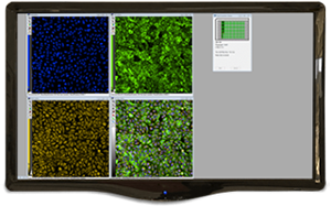
MetaXpress High-Content Image Acquisition and Analysis Software

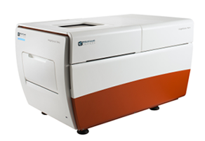
ImageXpress Nano Automated Imaging System

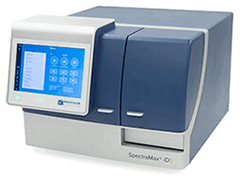
SpectraMax iD3 and iD5 Multi-Mode Microplate Readers
Products Used
The ImageXpress® Nano Automated Imaging System features a long life, solid state, light engine, and optics to reliably deliver the right assay sensitivity. Capture fine details of a variety of cellular and subcellular assays with this powerful and flexible fluorescent microscopy solution. The system includes MetaXpress® High-Content Image Acquisition and Analysis Software with tools for 2D and 3D imaging and time lapse analysis, as well as a range of needs from ease-of-use through to proprietary assay design.
The SpectraMax® iD3 and iD5 Multi-Mode Microplate Readers measure absorbance, fluorescence, and luminescence. In addition, the iD5 reader measures TRF and FP and can be expanded to include TR-FRET, HTRF®, BRET, dual luciferase reporter assays with injectors, and western blot detection. With optimized reagents, option to operate the readers using the touchscreen or a computer, and industry-leading SoftMax® Pro Software, the readers provide an all access pass for breakthrough research.
The Results
“The ImageXpress Nano Automated Imaging System offers a good amount of flexibility in imaging a wide variety of samples ranging from 3D cell aggregates to whole cell and sub-cellular organelles. Contrary to conventional fluorescence microscopes, the [ImageXpress Nano] allows focusing across different planes of a 3D structure (spheroid/ organoids). It also allows environmental control over the samples using preset humidity, CO2, and temperature control for time lapse experiments and live cell imaging features. The MetaXpress® High-Content Image Acquisition and Analysis Software enables imaging and analyses using predefined protocols for specific types of experiments while having the flexibility to set custom protocols. Overall, the ImageXpress Nano system is a great platform for multi-parametric and high throughput imaging and analyses.” - Dr. Shweta Bhatt, Chief Scientific Officer, Yashraj Biotechnology

Figure: Neurite outgrowth assay: Representative images of iPSCs-derived forebrain motor neurons with ß-tubulin (green) stain shown for the untreated and Taxol-treated cells. Forebrain motor neurons treated with compounds for three days and then fixed and stained with anti-ß-tubulin (TUJ-1).

Figure: Pluripotent iPS cells express pluripotency markers. (A), Oct-4 (B) Nanog (C) SSEA4. Nuclei counterstained with DAPI (blue).

Figure: Cytoskeleton disruption: Representative images of iPSC-derived hepatocytes with actin (green) stain shown for the untreated and Latrunculin-treated cells. Hepatocytes treated with compounds for three days and then fixed and stained with anti-Phalloidin-Actin.

Figure: TUNEL assay: Representative images of iPSC-derived cardiomyocytes with TUNEL (green) and Propidium Iodide (red) stain shown for the untreated and Doxorubicin-treated cells. Cardiomyocytes treated with compounds for three days and then fixed and stained with TUNEL and PI.

Figure: iPSCs-derived neural progenitor cells express neural progenitor markers PAX6 and Nestin. Nuclei counterstained with DAPI (blue).

Figure: A. iPSCs-derived forebrain motor neurons express neural markers Tuj1. B. iPSC-derived astrocytes express neural marker GFAP and C. iPSC-derived midbrain dopaminergic neurons express neural marker thymidine hydroxylase (TH). Nuclei counterstained with DAPI (blue).