- Home
- Products
- Cellular Imaging Systems
- Acquisition and Analysis Software
- MetaXpress High-Content Image Acquisition and Analysis Software
High-content image analysis software featuring time lapse analysis
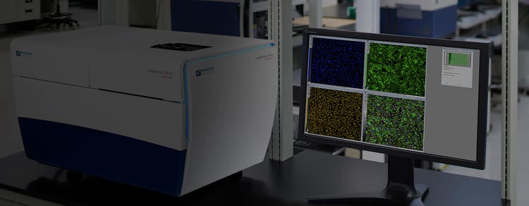
MetaXpress® High-Content Image Acquisition and Analysis Software is a comprehensive solution for high-content analysis featuring a tightly orchestrated and integrated workflow. The portfolio of application modules supports a range of needs from ease-of-use through to proprietary assay design. The software includes powerful and elegant tools for 2D and 3D imaging such as time lapse analysis. With the assistance of our acquisition setup wizard, you’ll be generating data in minutes.
-
Acquire and analyze in 2D and 3D
MetaXpress software is a platform for 2D and 3D image acquisition and analysis. The 3D Image Analysis Module can analyze spheroids, microtissues, cells in a 3D matrix, and small organisms.
-
Streamline image analysis
The modular toolbox allows for the quick setup of hundreds of routinely used high-content assays. Choose from our optional selections of turnkey application modules for greater convenience.
-
Design custom analyses easily
Quickly create multi-step routines with the Custom Module Editor. It enables advanced analyses such as identifying objects within objects, and creating morphometric classifiers for shape analysis.

Introducing the MetaXpress 3D Analysis Toolkit
Features
-
Z-stacks and 3D volumetric analysis
The integrated acquisition and analysis application modules for 3D cell models simplify high-throughput quantification of 3D structures with volume, intensity, and distance measurements.
-
Fast acquisition and analysis
Easily captures fast biological functions with our patented fast frame rate that can capture 50 frames per second. A 1-color, 384-well plate analysis can be performed in as little as six minutes.
-
Intelligent segmentation
Application modules feature turnkey image segmentation for label-free and fluorescently labeled images. The Custom Module Editor offers advanced tools for improved segmentation.
-
Scalable parallel processing
MetaXpress PowerCore™ Software runs analysis routines in minutes. This optional module leverages the power of parallel processing to accelerate analysis.
-
Data management solution
MDCStore™ Data Manager, included with MetaXpress software, organizes images, experimental information, and data analysis. It enables seamless manipulation of complex sets of images and metadata.
-
Advanced informatics
AcuityXpress™ High-Content Informatics Software is an optional enterprise level platform for advanced data mining, analysis, statistics, and visualization. It shares the MDCStore database for seamless integration.
Turnkey solutions for most common analysis routines
MetaXpress Application Modules
Expedite image analysis and data processing using our MetaXpress software with application modules that allow you to quickly and easily analyze your data:
- Micronuclei
- Neurite Outgrowth
- Angiogenesis Tube Formation
- Mitotic Index
- Plus many more applications...
Latest Resources
MetaXpress Software Application Modules
Set up hundreds of routinely used HCS assays whether you’re a novice or an expert. MetaXpress software application modules give you an interactive environment to quantify 2D and 3D measurements, including size, intensities, and distance.
-
3D Image Analysis Module
Complex assays and three-dimensional cell models better represent tissue biology and cell interactions, making them more relevant for many toxicity and drug screening assays. The ability to use 3D models in a high-throughput, high-content format is a significant benefit for scientists. The 3D Image Analysis Module for MetaXpress® Software is designed to analyze spheroids, microtissues, cells in a 3D matrix, and small organisms that are acquired as a stack of z-planes.
Angiogenesis Tube Formation
Angiogenesis Tube Formation Application Module facilitates the analysis of tube formation, a model experimental system for angiogenesis. The promotion or inhibition of blood vessel formation is one focus for cancer, diabetes and other vascular disease research.
-
Cell Cycle
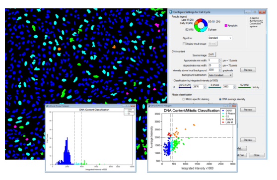
Cell Cycle Application Module classifies and quantifies cells in various stages of the cell cycle to investigate cell cycle progression. In healthy non-cancerous cells, challenges with DNA damage, hypoxia, metabolic changes or spindle disruption result in the triggering of checkpoints and cell cycle arrest. Cancerous cells commonly lose checkpoints and divide uncontrollably, even in challenging conditions. With the appropriate tools, researchers can screen for drugs that cause cell cycle arrest or cell death in cancerous cells.
Cell Health
Cell Health Application Module classifies cells states in viable, early apoptosis, late apoptosis or necrosis.
-
Cell Proliferation High-Throughput
Cell Proliferation HT Application Module is a high throughput version of the Count Nuclei, (about 1 minute for 96-well plate at one site per well without the MetaXpress® PowerCore™ Software) with a simplified detection algorithm, which provides site summary data only (not cell-by-cell) and does not save segmentation overlays to the MDCStore Database.
Cell Scoring
Cell Scoring Application Module is a general and flexible solution to identify subpopulations of cells tagged with a second fluorescent probe and is ideal for examining transfection efficiencies, pathway activation (kinases) or adipogenesis.
-
Count Nuclei
Count Nuclei Application Modules automate accurate counting of nuclei for most types of cells and are ideal for the study of cell proliferation, cell counting or cell migration. This module counts nuclei even when the background is uneven, providing superior segmentation compared to simple thresholding.
Custom Module Editor
The Custom Module Editor in version 5 of MetaXpress® Software is a flexible, interactive environment where users can create and test a template for image analysis. Galleries of processing options, morphology filters, application modules, and segmentation methods are available for users to construct a sequence of analysis steps. The new Click-to-Find Tool enables easy object identification and auto-populates step card parameters. Once created, custom modules can be easily shared and re-used to analyze other image sets.
-
Granularity and Transfluor
Granularity and Transfluor Application Module facilitate the analysis of punctate structures such as the one observed during clustering of target molecules for receptor internalization or within the nucleus or even the punctate patterns of mitochondria or other organelles.
Live/Dead
Live Dead Application Module is compatible with commercially available Live/Dead assay kits designed to study cell proliferation or death. Common applications monitor cell proliferation associated with cancer or cell death due to neuromuscular diseases such as Alzheimer's and Parkinson's Disease or in response to cytotoxic and apoptotic events.
-
Micronuclei
Micronuclei Application Module classifies cells based on nuclear morphology for genotoxicity applications but is also ideal for cell health or multi-nucleated cell populations with additional markers for analysis of apoptosis, necrosis or to distinguish any small structure next to a large one such as a yeast bud.
Mitotic Index
Mitotic Index Application Module is designed for the quantitative discrimination of mitotic and interphase cells, a critical tool for oncology drug discovery programs, providing insight into potential anti-cancer therapeutics that rely on arresting mitosis in cancerous cells to prevent uncontrolled proliferation.
-
Monopole Detection
Monopole Detection Application Module monitors the disruption of the formation of bipolar spindles, a successful mechanism used by drugs such monastol to stop the progression of cancerous cells through mitosis. The module quantifies mitotic cells with monopolar or bipolar spindles in cells stained with a DNA probe and a microtubule probe.
Multi-Wavelength Cell Scoring
Multi-Wavelength Cell Scoring Application Module enhances the functionality of the Cell Scoring Application Module to include additional wavelengths to score populations of cells based on numerous labels.
-
Multi-Wavelength Translocation
The Multi-Wavelength Translocation Application Module lets you analyze the translocation of up to 6 probes simultaneously (or the same probes with various settings), in addition to the nuclear stain.
Neurite Outgrowth
Neurite Outgrowth Application Module is designed to measure and characterize outgrowths, the extension of processes from the cell body, which are natural parts of neuronal development. Inhibition or stimulation of neurite outgrowth is implicated in a broad range of neurological disorders or injuries including stroke, Parkinson's disease, Alzheimer's disease, and spinal cord injuries.
-
Nuclear Translocation High-Throughput
Nuclear Translocation HT Application Module is for the fast analysis of translocation in and out of the nuclear compartment (about 1 minute for 96-well plate at one site per well without MetaXpress PowerCore Software) with a simplified detection algorithm, which provides site summary data only (not cell-by-cell) and does not save segmentation overlays to the MDCStore Database.
Toxicology
Toxicology is the study of adverse effects of natural or man-made chemicals on living organism. It is a growing concern in our world today as we are exposed to more and more chemicals, both in our environment and in the products we use.
-
Transfluor High-Throughput
Transfluor HT Application Module is a high throughput version of the Granularity Application Module for fast screening (about 1 minute for 96-well plate at one site per well without the MetaXpress PowerCore Software) with a simplified detection algorithm, which provides site summary data only (not cell-by-cell) and does not save segmentation overlays to the MDCStore Database.
Translocation and Translocation-Enhanced
Translocation and Translocation-Enhanced Application Modules quantify the correlation between probes and compartments. Translocation is ideal for studying general cellular component movement from the cytoplasm to the nucleus with a minimum number of adjustments. While Translocation-Enhanced offers increased functionality with finer definition of regions for translocation and the option to study translocation in non-nuclear compartments such as mitochondria.
Resources of MetaXpress High-Content Image Acquisition and Analysis Software
Application Note
Monitoring of T-cell invasions assay using a 3D spheroid model
T-cell therapies are designed to help our immune system eliminate cancer cells. Those include CAR T-cells (Chimeric Antigen Receptor engineered T-cells), tumor infiltrating lymphocytes…
Brochure
BioAssemblyBot 400 (BAB400): more than a bioprinter
The BioAssemblyBot® 400 is an intelligent robot used by life scientists to build 3D model systems with increased throughput and precision, alleviating major concerns mentioned above.…
Application Note
Automated dispensing, monitoring, and assay development of hydrogel-based cellular models
3D cellular models that more accurately represent various microenvironments are incredibly important for accurate drug screening and disease modeling.
Scientific Poster
Automation for organoid assays – An integrated system with high-content imaging
Three-dimensional (3D) cell models that represent various tissues are being successfully used in drug discovery and disease modeling to study complex biological effects and tissue ar…
Blog
Stem Cell Science and Regenerative Medicine – Technology and Methods Presented at ISSCR 2022
It was another great year at ISSCR 2022! Leaders from across the globe came together to discuss new technologies, share insights, and explore the newest breakthroughs in stem cell…
Application Note
Neurite outgrowth analysis in 3D reconstructed human brain micro-tissues
Organotypic 3D cultures resemble native 3D tissue architecture and are believed more representative of real tissues than their 2D (monolayer) counterparts, thus providing higher in…
Scientific Poster
Structural organization in 3D human iPSC-derived cardiac tri-culture microtissues
The human heart is a complex organ providing highly regulated processes of moving blood through the body. The adult human ventricle is comprised of cardiomyocytes, endothelial cells,…
Scientific Poster
Automated monitoring of development and activity analysis of iPSC-derived 3D cerebral organoids
The culture of complex organoids such as the cerebral organoid is a rapidly developing technology that has immense potential in areas of developmental neurobiology and neurodegenerat…
Scientific Poster
Monitoring growth and characterization of compound effects in 3D intestinal organoids by high content imaging
3D cell models representing various tissues were successfully used for studying complex biological effects and tissue architecture, however, the complexity of 3D models remains a hur…
Application Note
Disease modeling methods with cancer patient-derived organoids
The use of patient-derived tumor tissue has transformed the field of drug and target discovery research, providing a translational tool and physiologically relevant system to evaluate…
eBook
The essential guide to organoids in drug discovery
Three-dimensional (3D) organoid development is one of the most important advancements in drug discovery research to date.
Application Note
3D Immune Cell Migration
Cell migration is an essential process for many biological phenomena such as early embryonic development and immune cell response. During inflammation, the recruitment of T cells…
Scientific Poster
Phenotypic profiling based on the Cell Painting assay
Multiparametric high-content screening approaches, such as the Cell Painting assay, are increasingly being used in many applications ranging from drug discovery programs to functiona…
Scientific Poster
Automation of the organ-on a chip assay: automated culture, imaging and analysis of angiogenesis
There is a critical need for biological model systems that better resemble human biology. Three-dimensional (3D) cell models and organ-on-a-chip (OoC) structures representing various…
Scientific Poster
Intestinal organoids for automated screening assays. High content imaging and analysis of organoid morphology
Here we describe a workflow for automation of organoid culture. The automated method utilizes an integrated work-cell, consisting of several instruments providing automated cell cult…
Blog
Engineering Next-gen Organoids with Automated Lab Workflows at #SLAS2022
SLAS2022, the Society for Lab Automation and Screening conference offered another exciting year for learning about innovative laboratory technologies. Whether you attended in-person…
Blog
Enabling 3D High-Content Imaging and Analysis on the Organ-on-a-Chip Platform
Imagine having the ability to mimic the human biological environment for disease modeling and drug screening and doing so in a micro-scale system. With the development of organ-on-a-…
Scientific Poster
Automation of 3D organoid culture workflow with deep-learning based image analysis
3D cell culture as a model system is increasingly popular because it recapitulates the in vivo microenvironment better than 2D cell cultures. Organoids have the capacity for stable d…
Scientific Poster
Automation and high content imaging of 3D triple-negative breast cancer patient-derived
Triple negative breast cancer is a clinically aggressive tumor subtype, with high rates of metastasis, recurrence, and drug resistance. Currently there are no clinically approved sma…
Customer Breakthrough
Yashraj Biotechnology leverages the ImageXpress Nano to substantially improve their multi-parametric imaging and analyses
Unexpected toxicity in areas such as cardiotoxicity, hepatotoxicity, and neurotoxicity is a serious complication of clinical therapy and one of the key causes for failure of promisin…
Customer Breakthrough
Visikol team develops 3D cell culture models to simulate the NASH and NAFLD disease progression process using ImageXpress Micro Confocal High-Content Imaging System and MetaXpress software
In the Non-Alcoholic Steatohepatitis (NASH) and Nonalcoholic Fatty Liver Disease (NAFLD) space, there has been a strong need for an in vitro model that allows researchers to better s…
Blog
Innovation at Molecular Devices: Updates in Automated, High-Content Imaging
From customer feedback to workflow improvements The path to understanding complex biological processes and diseases is paved with a lot of challenges. As the desired level of…
Blog
Stem cell science insights and breakthroughs presented at #ISSCR2021
If you didn't get a chance to visit us at our poster sessions during ISSCR 2021, don’t fret. We've gathered all our sessions right here for you. The ISSCR Annual Meeting brought…
Publications
Project Profile: Organoid Innovation Center
Lab Manager speaks to Dan O’Connor, vice president, drug discovery, Molecular Devices, about the company’s Organoid Innovation Center in San Jose, CA. The center is 180 sq. ft., with…
Publications
Organs-on-Chips: Expand the Boundaries of In Vitro Testing
Humans began culturing animal cells and tissues as early as the late 19th century, when Wilhelm Roux first showed that chick embryos could be cultured in saline solution for a few da…
Blog
Tips for running a successful live cell imaging experiment
There have been significant advancements in microscopy and camera technology, as well as advancements in technologies for labeling molecules of interest over the past decade. These…
Blog
Overcome the challenges of high-throughput 3D imaging
Thanks to recent advances in imaging technologies, we are now able to observe and analyze complex cellular networks in three dimensions. Through 3D imaging, we can acquire and…
Publications
Shaping the Future of Organoid Research
Molecular Devices, a provider of innovative life science technology, recently unveiled a brand new, first-of-its-kind Organoid Innovation Center. Situated at the company’s global hea…
Application Note
Lung organoids as an assay model for in vitro assessment of toxicity effects by 3D high-content imaging and analysis
Organoid models have increasingly gained popularity in biologic research and screening to recapitulate complexity of real tissues. To model the in vivo human lungs, we have cultured…
Blog
Overcome the challenges of high-content cell analysis through AI/machine learning
Artificial intelligence (AI) is finding its way into many aspects of modern life, from autonomous vehicles to voice-powered personal assistants, and even the creation of art. But it…
Publications
Disease Modeling with 3D Cell-Based Assays Using a Novel Flowchip System and High-Content Imaging
There is an increasing interest in using three-dimensional (3D) cell structures for modeling tumors, organs, and tissue to accelerate translational research. We describe here a novel…
Blog
Get to Know our Field Applications Scientist: Cheryl Bell
Cheryl Bell talks about making the transition from manual microscopy to automated microscopy Our Field Applications Scientist, Cheryl Bell, was recently featured in a Women in…
Scientific Poster
Automated 3D cell-based assays using a novel flow chip system and high-content imaging
There is an increasing interest in using three-dimensional (3D) cell structures for modeling tumors, organs, and tissue to accelerate translation research. Significant progress has b…
Blog
How Cell Painting is making its mark on drug discovery
Have you ever heard the old adage, “A picture is worth a thousand words?” When it comes to Cell Painting, this saying is especially true. Cell Painting is a high-content,…
eBook
Cellular Imaging Insights
Gain insights and expedite studies for 2D and 3D cellular structures using automated cellular imaging.
Blog
Life sciences technology predictions for 2021
For over 30 years, Molecular Devices has been at the forefront of technological advances which have contributed to significant scientific breakthroughs. To kick off the new year, we…
Blog
7 tips for optimizing your 3D cell imaging and analysis workflow
As the field of cell biology moves toward increasing complexity of assays, 3D cell systems have proven to provide more physiologically relevant information as compared to…
Blog
Water immersion technology and high-content imaging: A closer look
Three-dimensional (3D) cellular assays have emerged as a valuable tool in drug discovery and biological research, as they closely mimic in vivo environments and are shown to…
Blog
Get to know our Field Applications Scientist: Kayla Hill
Kayla Hill discusses the latest trends in cellular imaging We recently hosted a webinar with our Field Applications Scientist, Kayla Hill, PhD, who explored high-content analysis…
Application Note
High-throughput screening of 3D cell cultures with multiple high density scaffold-free spheroids for cancer toxicity studies
3D spheroid models for cancer research are gaining popularity because they better mimic the in vivo tissue architecture, gene expression and metabolic profile of tumors compared to…
Application Note
3D image analysis and characterization of angiogenesis in organ-on-a-chip model
Angiogenesis is the physiological process of formation and remodeling of new blood vessels and capillaries from pre-existing blood vessels. This can be achieved through endothelial…
Scientific Poster
Automated Long-term 3D Cell-Based Toxicity Studies Using a Flowchip System
There is an increasing interest in using three-dimensional (3D) cell structures for modeling tumors, organs, and tissue to accelerate translation research.1 Significant progress has…
Publications
3D Microscopy Keeps Getting Faster, Smarter, Leaner
With each passing day, continuous advancements are being made in the studies that cutting-edge instruments can perform and in the complexity of the biological samples that are being…
Publications
From 3D high throughput to simple automated imaging: Cellular imaging within reach of every laboratory
OUR ABILITY to image cells has come a long way since the pioneering days of Galileo Galilei and Antonie van Leeuwenhoek. The multiple imaging techniques available today range from si…
Publications
Revolutionizing early drug discovery for immuno-oncology and neurodegenerative disease modeling
The development of 3D cellular models of disease has enabled researchers to better recapitulate in vivo cell environments. As 3D models become more prevalent in the drug discovery in…
Publications
LabTube Meets Molecular Devices & MIMETAS
Molecular Devices partners with MIMETAS to scale up organoid screening—helping researchers go from 2D to 3D phenotypic relevant organoid screening. At SLAS 2020, they featured the Im…
Publications
The importance of the cellular microenvironment in the development of more efficient drugs against complex diseases
The biotechnological start-up is focused on in vitro preclinical research and development, with a specialization in automated cellular imaging: high-content analysis (HCA) and high-c…
Application Note
High-throughput screening of single-cell contractile force using FLECS Technology
The ability of mammalian cells to generate mechanical forces—to push, pull, or squeeze—is an intrinsic capability that is used by cells both individually and together as tissue to…
Customer Breakthrough
HCS Pharma uses ImageXpress Micro Confocal Systems for phenotypic screening of physiologically relevant 3D cell models
HCS Pharma is a biotechnology start-up focused on in vitro preclinical research and development. The company specializes in multi-parametric cell imaging assays using High-C…
Publications
Rising growth of the global high‑content screening software and services market
Developments in informatics solutions and high-content imaging instruments, increased focus on cell-based research, and rising government support of research and development for drug…
Application Note
Counting cells with the Transmitted Light Analysis Module in MetaXpress
Label-free cellular assays are required for a multitude of biological applications that monitor the cell number, proliferation, health, confluency, and cytotoxicity. In this…
Application Note
Analyze experimental results using curve fitting with MetaXpress 6.5 software
The ability to annotate your plate, plot your data into a graph, and produce a variety of curves and readouts including IC50 values and benchmark dose or concentration for your…
Customer Breakthrough
Gustave Roussy Cancer Center uses the ImageXpress Widefield Systems to assess mechanisms of cell stress and death in cancer treatment
The Kroemer Lab is interested in the molecular mechanisms of cellular stress and death. The mode of cellular stress and death that is evoked during cancer treatment has profound ther…
eBook
Acquire and analyze 3D images like a pro
There has been significant progress in the development of 3D models and techniques during the last few years. Methods include biodegradable scaffolds, organ-on-a chip structures, or…
Brochure
Imaging Solutions to Answer Your Biological Questions
Learn about our imaging family and which solution is best suited to answer your biological questions.
Brochure
MetaXpress High-Content Image Acquisition and Analysis Software
Capture high-resolution images, recognize and segment objects, calculate parameters, and then translate your information into meaningful data. At Molecular Devices, we’re inspired by…
Data Sheet
MetaXpress Software High-Throughput Application Modules
MetaXpress is high throughput module that accelerates image-based screening process & is used in conjunction by corresponding research modules for cell-by-cell data mining.
Data Sheet
MetaXpress Software Count Nuclei Application Module
Count Nuclei is designed to automate accurate counting of most types of cells. This is a user-friendly module to minimize set-up efforts & provides flexibility to customize the s…
Data Sheet
MetaXpress Software Multi-Wavelength Cell Scoring Application Module
Multi-Wavelength Cell scoring datasheet describes the MetaXpress App Module that makes a number of additional cell-by-cell measurements including per-wavelength count.
Data Sheet
MetaXpress Software Cell Scoring Application Module
Get an overview of Cell Scoring Application designed to identify two subpopulations of cells & to make a number of measurements including the number & percentage of cells.…
Data Sheet
MetaXpress Software Monopole Detection Application Module
The PDF defines Monopole Detection Module designed for quantitation of mitotic cells with monopolar or bipolar spindles.
Data Sheet
MetaXpress Software Cell Cycle Application Module
This Cell Cycle datasheet provides an overview of the MetaXpress App Module. It is designed for the quantitation of cell cycle stage for cells labeled with a DNA stain.
Data Sheet
MetaXpress Software Mitotic Index Application Module
Explore Mitotic Index datasheet to get information about MetaXpress App Module designed for quantitative discrimination of mitotic & interphase cells.
Flyer
Digital Confocal for MetaXpress
Get details about digital confocal option for ImageXpress Micro Imaging Systems that enhance image contrast during acquisition & allow decreasing exposure time to maintain assay…
Data Sheet
MetaXpress Software Micronuclei Application Module
Get details about MetaXpress Application Module Micronuclei which includes cell cycle analysis, cell health profiling, live/dead determination, and mitotic index calculation.
Data Sheet
MetaXpress Software Translocation Application Modules
The Translocation and Translocation-Enhanced Application Modules for MetaXpress® Software from Molecular Devices compliments the suite of application modules for the quantification o…
Data Sheet
MetaXpress Software Multi-Wavelength Translocation Application Module
Multi-Wavelength Translocation Module is designed for the combination, or multiplexing, of multiple translocating probes into a single assay. Explore this document to know more.
Data Sheet
MetaXpress Software Cell Health Application Module
This datasheet shows Cell-based assays that provide an efficient method of modeling toxicological effects of drugs & for discriminating anti-proliferative effects.
Data Sheet
MetaXpress Software Transfluor Application Module
The Transfluor Application Module for MetaXpress® Software from Molecular Devices is designed to facilitate the analysis of receptor internalization specifically for the Transfluor a…
Data Sheet
MetaXpress Software Live/Dead Application Module
This LiveDead datasheet describes MetaXpress App Module is designed to automate the accurate identification of cells after staining with a variety of live/dead staining kit.
Customer Breakthrough
SENS Research Foundation uses the ImageXpress Micro Widefield system to speed up cancer research 20 times
The Oncology team at the SENS Research Foundation seeks to uncover the role of telomere lengthening in cancer development. The Alternative Lengthening of Telomeres (ALT) pathway acco…
Application Note
High-content analysis of the various stages of apoptosis with the MetaXpress Cell Health Application Module
Cell-based assays provide an efficient method of modeling the toxicological effects of drugs and for discriminating anti-proliferative effects from induction of apoptosis or necrosis.…
Application Note
High-Content Screening of GPCR activation Using MetaXpress and AcuityXpress Software and the Transfluor Assay System
G-protein coupled receptors (GPCRs are the largest class of pharmaceutical targets and, as a result, assays for detecting GPCR receptor agonists and antagonists play a major role in…
Application Note
Custom Module Editor For MetaXpress High Content Image Acquisition and Analysis Software
The Custom Module Editor in version 5 of MetaXpress® Software is a flexible, interactive environment where users can create and test a template for image analysis. Galleries of…
Flyer
MetaXpress 6 Software
Reimagine your current workflow with MetaXpress® High-Content Image Acquisition and Analysis Software. The latest layout simplifies plate acquisition setup by consolidating key funct…
Application Note
MetaXpress Software Angiogenesis Application Module
Angiogenesis is the growth of new blood vessels and is an important natural process occurring in the body, both in health and disease. Angiogenesis occurs in the healthy body for…
Application Note
Intelligent time-lapse imaging to enhance live cell assay development
The ability to monitor responses in living cells over a specific period of time offers cell biologists several key advantages for assay development. For routine cellbased screening,…
Flyer
Neurite Outgrowth Application Module
Explore the Neurite Outgrowth Application Module for MetaXpress software, which is designed for the analysis of neurite outgrowth assays
Brochure
Innovative Solutions for Drug Discovery and Life Sciences Research
Molecular Devices is a leading provider of high-performance bioanalytical measurement solutions for life science research, pharmaceutical development, and biotherapeutic discovery. O…
Brochure
3D Image Analysis Module
Complex assays and three-dimensional cell models better represent tissue biology and cell interactions, making them more relevant for many toxicity and drug screening assays. The abi…
Publications
Phenotypic Characterization of Toxic Compound Effects on Liver Spheroids Derived from iPSC Using Confocal Imaging and Three-Dimensional Image Analysis
Cell models are becoming more complex to better mimic the in vivo environment and provide greater predictivity for compound efficacy and toxicity. There is an increasing interest in…
Publications
A Chemical-Biological Similarity-Based Grouping of Complex Substances as a Prototype Approach for Evaluating Chemical Alternatives
Comparative assessment of potential human health impacts is a critical step in evaluating both chemical alternatives and existing products on the market. Most alternatives assessment…
Publications
High-Content Assay Multiplexing for Toxicity Screening in Induced Pluripotent Stem Cell-Derived Cardiomyocytes and Hepatocytes
Cell-based high-content screening (HCS) assays have become an increasingly attractive alternative to traditional in vitro and in vivo testing in pharmaceutical drug development and t…
Publications
High-Content Assays for Characterizing the Viability and Morphology of 3D Cancer Spheroid Cultures
There is an increasing interest in using three-dimensional (3D) spheroids for modeling cancer and tissue biology to accelerate translation research. Development of higher throughput…
Publications
High-Content Assays for Hepatotoxicity Using Induced Pluripotent Stem Cell–Derived Cells
Abstract Development of predictive in vitro assays for early toxicity evaluation is extremely important for improving the drug development process and reducing drug attrition rates d…


Ananda Neurite Untangled

Leverage automated, end-to-end workflows to enable complex organoid assays

Level Up your 3D Cell Culture: From Research to High-Throughput

Enhance high-content 3D biology imaging with automated sample preparation

Capturing the complexity of 3D biology: Organoids for disease modelling and toxicity research

Imaging and Analysis of a 3D Airway Organoid Model

Immunology and Vaccine Development Workflow

Hybridoma Workflow

Plate annotation and curve fitting in MetaXpress

Quickstart Guide: Plate acquisition on the ImageXpress Micro Confocal using MetaXpress

Quickstart Guide: Review images on the ImageXpress Micro Confocal using MetaXpress

Basic workflow from image acquisition to analysis on the ImageXpress Micro Confocal using MetaXpress

High-Content Screening for Identifying miRNAs

Identification of Selective Inhibitors of the STAT3 Signaling Pathway

Oliver Kepp and Jayne Hesley - Hallmarks of Cancer - Detect and Quantify Cell Death Signatures with High Content Imaging

Multidimensional High-Throughput Imaging with the New ImageXpress Micro Confocal System

Pushing the Boundaries of High Content Screening

Preparing Assays for Genome-wide RNAi Screens Using High Content Microscopy

Multiplexed High Content Hepatoxicity Assays Using iPSC-Derived Hepatocytes

High Content Imaging Analysis of Cell Sheet Morphogenesis Utilizing 𝘪𝘯 𝘷𝘪𝘵𝘳𝘰 Tissue Models

Simple and Flexible High Content Imaging Enabling Quantitation of Complex Biological Events

Contemporary Automation and High Content Imaging Tools for Screening Stem Cell-Derived Cardiomyocytes

Implement High-Throughput 3D Image Analysis for Samples from Subcellular Structures to Spheroids

Live Cell Imaging to Investigate the Regulation of Cell Division Timing

Using High-throughput RNA Screening to Identify Host Factors That Impact Viral Infection

Application of HCA Tools for Antibody Drug Discovery

Setting up 3D Spheroid Assays Using High-Content Imaging

Emerging Induced Pluripotent Stem Cell Applications in Drug Discovery
Number of Citations*: 2,810
Latest Citations: For a complete list, please click here .
*Source: https://scholar.google.com/
- Dated: Jun 15, 2022Publication Name: American Association for Cancer Research (AACR) Journals
Abstract 192: Ex vivo screening and analysis of novel effective treatments for ovarian cancer
Purpose: Ovarian cancer is the most lethal gynecological cancer. Histologically, about 90% of the ovarian cancers have epithelial origin and more than 75% are characterized as high-grade serous ovarian cancer (HGSOC). View morePurpose: Ovarian cancer is the most lethal gynecological cancer. Histologically, about 90% of the ovarian cancers have epithelial origin and more than 75% are characterized as high-grade serous ovarian cancer (HGSOC).
Contributors: Aikaterini Skorda; Kaisa Huhtinen; Wojciech Senkowski; Krister Wennerberg; Sampsa Hautaniemi; Johanna Hynninen; Tuula Kallunki
Go to article - Dated: Dec 07, 2012Publication Name: PLOS ONE
Analyzing Illumina Gene Expression Microarray Data from Different Tissues: Methodological Aspects of Data Analysis in the MetaXpress Consortium
Microarray profiling of gene expression is widely applied in molecular biology and functional genomics. Experimental and technical variations make meta-analysis of different studies challenging. In a total of 3358 samples, all from German population-based cohorts, we investigated the effect of data preprocessing and the variability due to sample… View moreMicroarray profiling of gene expression is widely applied in molecular biology and functional genomics. Experimental and technical variations make meta-analysis of different studies challenging. In a total of 3358 samples, all from German population-based cohorts, we investigated the effect of data preprocessing and the variability due to sample processing in whole blood cell and blood monocyte gene expression data, measured on the Illumina HumanHT-12 v3 BeadChip array
Contributors: Claudia Schurmann, Katharina Heim, Arne Schillert, Stefan Blankenberg, Maren Carstensen, Marcus Dörr, Karlhans Endlich, Stephan B. Felix, Christian Gieger, Harald Grallert, Christian Herder, Wolfgang Hoffmann, Georg Homuth, Andreas Ziegler
Go to article - Dated: Oct 25, 2011Publication Name: Springer Nature
Immunohistochemical characterisation of cells co-producing insulin and glucagon in the developing human pancreas
In adult human islets, insulin and glucagon production is largely restricted to individual cell populations. The production of these hormones is less segregated during development and during the differentiation of human pluripotent stem cells towards pancreatic lineages. We therefore sought to characterise the transcription factor profile of these… View moreIn adult human islets, insulin and glucagon production is largely restricted to individual cell populations. The production of these hormones is less segregated during development and during the differentiation of human pluripotent stem cells towards pancreatic lineages. We therefore sought to characterise the transcription factor profile of these cells that co-produce insulin and glucagon in the developing human pancreas, and thus to gain insight into their potential fate during normal pancreas development.
- Dated: Feb 15, 2008Publication Name: Journal of Neuroscience Methods
NeuriteTracer: A novel ImageJ plugin for automated quantification of neurite outgrowth
In vitro assays to measure neuronal growth are a fundamental tool used by many neurobiologists studying neuronal development and regeneration. The quantification of these assays requires accurate measurements of neurite length and neuronal cell numbers in neuronal cultures. Generally, these measurements are obtained through labor-intensive manual… View moreIn vitro assays to measure neuronal growth are a fundamental tool used by many neurobiologists studying neuronal development and regeneration. The quantification of these assays requires accurate measurements of neurite length and neuronal cell numbers in neuronal cultures. Generally, these measurements are obtained through labor-intensive manual or semi-manual tracing of images. To automate these measurements, we have written NeuriteTracer, a neurite tracing plugin for the freely available image-processing program ImageJ. The plugin analyzes fluorescence microscopy images of neurites and nuclei of dissociated cultured neurons. Given user-defined thresholds, the plugin counts neuronal nuclei, and traces and measures neurite length. We find that NeuriteTracer accurately measures neurite outgrowth from cerebellar, DRG and hippocampal neurons. Values obtained by NeuriteTracer correlate strongly with those obtained by semi-manual tracing with NeuronJ and by using a sophisticated analysis package, MetaXpress. We reveal the utility of NeuriteTracer by demonstrating its ability to detect the neurite outgrowth promoting capacity of the rho kinase inhibitor Y-27632. Our plugin is an attractive alternative to existing tracing tools because it is fully automated and ready for use within a freely accessible imaging program.
Related Products & Services of MetaXpress High-Content Image Acquisition and Analysis Software

Advanced Cloud-Based Analytics with StratoMineR
Increase efficiency and throughput with cloud-based advanced analytics

Cellular Imaging Systems
Transform your incredible images into captivating results
Featured Applications

3D Image Analysis Module
Complex assays and three-dimensional cell models better represent tissue biology and cell…

Angiogenesis Tube Formation
Angiogenesis Tube Formation Application Module facilitates the analysis of tube formation, a model…

Cell Cycle
Cell Cycle Application Module classifies and quantifies cells in various stages of the cell cycle…

Cell Health
Cell Health Application Module classifies cells states in viable, early apoptosis, late apoptosis…

Cell Proliferation High-Throughput
Cell Proliferation HT Application Module is a high throughput version of the Count Nuclei, (about 1…

Cell Scoring
Cell Scoring Application Module is a general and flexible solution to identify subpopulations of…

Count Nuclei
Count Nuclei Application Modules automate accurate counting of nuclei for most types of cells and…

Custom Module Editor
The Custom Module Editor in version 5 of MetaXpress® Software is a flexible, interactive…

Granularity and Transfluor
Granularity and Transfluor Application Module facilitate the analysis of punctate structures such…

Live/Dead
Live Dead Application Module is compatible with commercially available Live/Dead assay kits…

Micronuclei
Micronuclei Application Module classifies cells based on nuclear morphology for genotoxicity…

Mitotic Index
Mitotic Index Application Module is designed for the quantitative discrimination of mitotic and…

Monopole Detection
Monopole Detection Application Module monitors the disruption of the formation of bipolar spindles…

Multi-Wavelength Cell Scoring
Multi-Wavelength Cell Scoring Application Module enhances the functionality of the Cell Scoring…

Multi-Wavelength Translocation
The Multi-Wavelength Translocation Application Module lets you analyze the translocation of up to 6…

Neurite Outgrowth
Neurite Outgrowth Application Module is designed to measure and characterize outgrowths, the…

Nuclear Translocation High-Throughput
Nuclear Translocation HT Application Module is for the fast analysis of translocation in and out of…

Toxicology
Toxicology is the study of adverse effects of natural or man-made chemicals on living organism. It…

Transfluor High-Throughput
Transfluor HT Application Module is a high throughput version of the Granularity Application Module…

Translocation and Translocation-Enhanced
Translocation and Translocation-Enhanced Application Modules quantify the correlation between…
Customer Breakthrough


SUCCESS STORY
Yashraj Biotechnology leverages the ImageXpress Nano to substantially improve their multi-parametric imaging and analyses
How can we help advance your next big discovery?
Our highly-qualified teams are on the frontlines with our customers, conducting remote or on-site product demonstrations, webinars, and more to help you solve your tough research challenges. How can we help you today?
I’d like to…
How can we help advance your next big discovery?
Our highly-qualified teams are on the frontlines with our customers, conducting remote or on-site product demonstrations, webinars, and more to help you solve your tough research challenges. How can we help you today?
I’d like to…