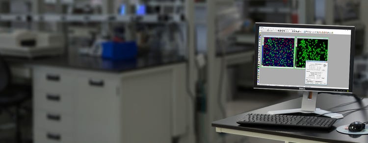
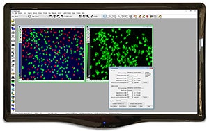
The MetaMorph® Microscopy Automation and Image Analysis Software automates acquisition, device control, and image analysis. It easily integrates dissimilar fluorescent microscope hardware and peripherals into a single custom workstation. The software offers many user-friendly application modules for biology-specific analysis. Two complementary packages, MetaFluor® software for fluorescence ratio imaging, and MetaVue® software for basic image acquisition and processing, are included in the portfolio.
The drug discovery landscape is shifting, with more scientists centering cell line development, disease models, and high-throughput screening methods around physiologically-relevant 3D cell models. The reason for this is clear: Using cellular model systems in research that closely mimic patient disease states or human organs can bring life-saving therapeutics to market – faster.
Toxicology is the study of adverse effects of natural or man-made chemicals on living organism. It is a growing concern in our world today as we are exposed to more and more chemicals, both in our environment and in the products we use.
Customer Breakthrough
The main fields of research for the team at the Laboratory of Mucosal Barrier Pathobiology, Department of Pathology, Brigham and Women’s Hospital, Harvard Medical School are epitheli…
Blog
There have been significant advancements in microscopy and camera technology, as well as advancements in technologies for labeling molecules of interest over the past decade. These…
Blog
Artificial intelligence (AI) is finding its way into many aspects of modern life, from autonomous vehicles to voice-powered personal assistants, and even the creation of art. But it…
Scientific Poster
There is an increasing interest in using three-dimensional (3D) cell structures for modeling tumors, organs, and tissue to accelerate translation research. Significant progress has b…
eBook
Gain insights and expedite studies for 2D and 3D cellular structures using automated cellular imaging.
Blog
For over 40 years, Molecular Devices has been at the forefront of technological advances which have contributed to significant scientific breakthroughs. To kick off the new year, we…
Blog
Kayla Hill discusses the latest trends in cellular imaging We recently hosted a webinar with our Field Applications Scientist, Kayla Hill, PhD, who explored high-content analysis…
Brochure
The MetaMorph® Microscopy Automation and Image Analysis Software from Molecular Devices plays a large role in this revolution. With its image acquisition, processing and analysis cap…
Customer Breakthrough
Dr. Jung-Chi Liao’s lab at Academia Sinica in Taiwan focuses on understanding the structure and function of proteins at the base of primary cilia in mammalian cells. Specifically, th…
Data Sheet
Explore Mitotic Index datasheet to get information about MetaXpress App Module designed for quantitative discrimination of mitotic & interphase cells.
Application Note
Neurons create connections via extensions of their cellular body called dendrites or “processes”. This biological phenomena is referred to as neurite outgrowth and is regulated by…
Application Note
Angiogenesis is the growth of new blood vessels and is an important natural process occurring in the body, both in health and disease. Angiogenesis occurs in the healthy body for…
Application Note
Bacterial screening is a popular method used for detecting recombinant bacteria in vector-based molecular cloning. It can also be used to measure specific protein expression and…
Brochure
The MetaFluor Imaging System is designed to display raw data, ratio image, graphs of intensities, ratios & ion concentrations, & ratiometric images such as brightfield or pha…
Brochure
Explore MetaMorph Microscopy & Imaging Software used to control experimental hardware, capture images, perform localization do calculations, and display super-resolution image de…
Application Note
Live cell research is increasingly in need of employing complementary data acquisition simultaneously to describe intracellular processes. Simultaneous recordings with both imaging and…
Immunology and Vaccine Development Workflow
Hybridoma Workflow
Resolving Molecular Organization and Dynamics Using Localization-Based Super-Resolution Microscopy
Real-Time Single-Molecule Based Super-Resolution Microscopy Reconstruction: Theoretical and Practical Insight
Latest Citations: For a complete list, please click here .
*Source: https://scholar.google.com/
Catalytic nanoparticles represent a potential clinical approach to replace or correct aberrant enzymatic activities in patients. Several diseases, including many blinding eye diseases, are promoted by excessive oxidant stress due to reactive oxygen species (ROS). Cerium oxide and platinum nanoparticles represent two potentially therapeutic nanoparticles that de-toxify ROS. In the present study, we directly compare these two classes of catalytic nanoparticles. Cerium oxide and platinum nanoparticles were found to be 16 ± 2.4 and 1.9 ± 0.2 nm in diameter, respectively. Using surface plasmon-enhanced microscopy, we find that these nanoparticles associate with cells. Furthermore, cerium oxide and platinum nanoparticles demonstrated superoxide dismutase catalytic activity, but did not promote hemolytic or cytolytic pathways in living cells. Importantly, both cerium oxide and platinum nanoparticles reduce oxidant-mediated apoptosis in target cells as judged by the activation of caspase 3. The ability to diminish apoptosis may contribute to maintaining healthy tissues.
Automated image-based and biochemical assays have greatly increased throughput for quantifying cell numbers in in vitro studies. However, it has been more difficult to automate the counting of specific cell types with complex morphologies in mixed cell cultures. We have developed a fully automated, fast, accurate and objective method for the quantification of primary human GFAP-positive astrocytes and CD45-positive microglia from images of mixed cell populations. This method, called the complex cell count (CCC) assay, utilizes a combination of image processing and analysis operations from MetaMorph™ (Version 6.2.6, Molecular Devices). The CCC assay consists of four main aspects: image processing with a unique combination of morphology filters; digital thresholding; integrated morphometry analysis; and a configuration of object standards. The time needed to analyze each image is 1.82 s. Significant correlations have been consistently achieved between the data obtained from CCC analysis and manual cell counts. This assay can quickly and accurately quantify the number of human astrocytes and microglia in mixed cell culture and can be applied to quantifying a range of other cells/objects with complex morphology in neuroscience research.
How a naive human neuroepithelial cell becomes an electrophysiologically active neuron remains unknown. Here, we describe the early physiological development of neurons differentiating from naive human embryonic stem (hES) cells. We found that differentiating neuronal cells progressively decrease their resting membrane potential, gain characteristic Na+ and K+ currents, and fire mature action potentials by 7 weeks of differentiation.