
Research team reduces scan times by 88% using MetaMorph Microscopy Automation and Image Analysis Software
COMPANY/UNIVERSITY
Laboratory of Mucosal Barrier Pathobiology, Department of Pathology, Brigham and Women’s Hospital, Harvard Medical School
TEAM MEMBERS
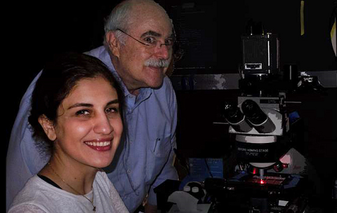
Jerrold R. Turner, M.D., Ph.D. , Professor of Pathology and Medicine
Shabnam Abtahi, MD Postdoctoral Research Fellow
PRODUCTS USED
MetaMorph® Microscopy Automation and Image Analysis Software
The Challenge
The solution
With the help of Molecular Devices, Dr. Turner’s team developed a set of custom journals incorporating the ScanSlide module that work together and are accessed via a unified taskbar that guides the user. This protocol allows:
- automated scanning of multiple regions of interest
- assisted ROI selection
- customizable, reliable software-based focusing
- simple, semi-automated and rapid review of preview images
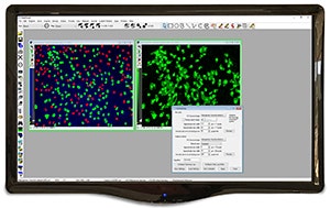
- automated stitching of high-quality images
As a result, Dr. Turner’s team can now complete a 20X objective scan of a slide with forty 3 mm tissue cores stained in five channels in less than 4 hours. Dr. Turner explains, “This compares favorably with microscope-based commercial systems. Because the system is run by MetaMorph, it allows complete flexibility in modifying any aspect of the system, including imaging modality, objectives used, illumination sources, filters, and cameras. For example, we recently added a switchable light source (Lumencore Aura III), motorized emission filter wheel (Ludl), and multichannel dichroic (Semrock) to eliminate the time lost while our microscope changed filter cubes (\~1 second per change). This cut another hour off our imaging time.”
MetaMorph Microscopy Automation and Image Analysis Software [Discontinued]
Products Used
MetaMorph® Microscopy Automation & Image Analysis Software is the industry standard for automated microscope acquisition, device control, and image analysis, bringing microscopists greater understanding of cell morphology, function, and behavior for over 25 years. It is the ideal "glue" for easily integrating dissimilar fluorescent microscope hardware and peripherals into a single custom workstation, while providing all the tools needed to perform meaningful analysis of acquired images. The software offers many user-friendly application modules for biology-specific analysis such as cell signaling.
The Results
- This system has allowed researchers to scan numerous tissue microarrays from experimental mice (Graham et al., 2019; Kuo et al., 2019; Nalle et al., 2019; Raju et al., 2020) and human biopsies (Turner, 2020)
- Over 5,000 scanned images have been compiled into the publicly accessible Atlas of Intestinal Transport (https://jrturnerlab.com/database-viewer/atlas-of-intestinal-transport/)
- The journals and Current Protocols publication (Abtahi et al, 2021) and an instructional video can be viewed and downloaded at https://jrturnerlab.com/publication/a-simple-method/