Automating the brain organoid culture process
Why automating brain organoid culture Is a game-changer for neuroscience research
“Hello, I’m Sandra Grund-Gröschke, a 3D Application Scientist here at Molecular Devices. Today, I’m excited to show you how automation is transforming the way scientists grow and study brain organoids.
https://vids.moleculardevices.com/watch/4oMgWVWge9oEPG9zP13BUn
With the CellXpress.ai Automated Cell Culture System, we’re combining intelligent automation, AI-driven monitoring, and our innovative rocking incubator to help researchers scale complex 3D biology—faster, more reproducibly, and with greater insight than ever before.
From stem cell cultivation to brain organoid differentiation, this system acts as a true lab partner—freeing scientists to focus on discovery while automation handles the routine.
Let’s take a closer look at how CellXpress.ai is redefining what’s possible in brain organoid research.”
Why are scientists using brain organoids for their neuroscience research?
Scientists are increasingly using brain organoids in neuroscience research because they provide a more accurate model of the human brain than traditional two-dimensional cell cultures. Derived from induced pluripotent stem cells (iPSCs), brain organoids are three-dimensional, miniaturized models that mimic both the architecture and functionality of human brain tissue.
Unlike flat 2D cultures that typically contain a single cell type, brain organoid models include multiple interacting cell types—such as neurons, astrocytes, and other supporting cells—that self-organize into structures resembling real brain regions. This complexity allows researchers to study cortical layering, regional specification, and neural activity in a way that closely mirrors actual human brain development.

Brain organoids replicate the structural and functional complexity of human brain tissue, making them superior to traditional 2D cultures.
Brain organoids are also opening doors to personalized medicine and a deeper understanding of the role the genome may play in disorders such as epilepsy. By generating organoids from the stem cells of patients with such disorders, there is now the potential to understand in far more detail what causes these conditions and how they might be treated.
Because of these advantages, brain organoids are now being used to:
- Model neurological diseases such as Alzheimer’s, Parkinson’s, and epilepsy
- Study human brain development at early stages
- Test potential therapeutic strategies in a controlled laboratory setting
By combining cellular diversity, structural organization, and functional activity, brain organoids are revolutionizing neuroscience research and offering insights that were not possible with simpler models.
Can brain organoid culture be automated?
Automating brain organoid culture addresses one of neuroscience research’s biggest challenges: complexity. Unlike manual methods that are time-consuming, variable, and prone to contamination, automation ensures consistent feeding, monitoring, and handling. This improves reproducibility, reduces errors, and frees researchers to focus on discovery instead of routine maintenance.
Challenges of Brain Organoid Culture: Why Manual Methods Fall Short
Despite their promise, brain organoids are notoriously difficult to culture manually, even compared to other organoids. Brain organoids require constant motion and regular feeding to develop properly.
Some of the challenges Dr. Sandra has observed include:
- A highly labor-intensive process that requires precise, consistent handling for periods often exceeding 100 days – which requires intervention during weekends and holidays.
- Manual processes introducing variability, which in turn impacts the accuracy and reliability of downstream assays.
- Increased risk of contamination, due to so much hands-on work over extended timelines.
Automated Brain Organoid Culture: A Scalable Solution for Neuroscience Research
Here’s how automation helps overcome the biggest challenges in brain cell culture:
- Feeding organoids automatically means they’re cared for on a set schedule, even on weekends and holidays. That consistency not only reduces human error but also leads to more reliable downstream assays.
- Scientists get their time back. With automated workflows, there’s no need to spend excessive amounts of time at the lab bench culturing organoids, come in outside of regular hours, or plan experiments around staff availability.
- Imaging becomes reliable and repeatable. Automated imaging systems take care of the routine image acquisition and analysis, freeing researchers from repetitive work.
- Contamination risks drop significantly. In a busy lab, it’s easy to mix up media or mishandle plates. Automated handling keeps every step standardized and secure.
Why Brain Organoid Automation Is Complex: Motion and Culture Requirements
So why has brain organoid automation not become mainstream until now? The answer lies in the unique and complex demands of brain organoid culture.
The need for constant motion
First, brain organoids must be kept in continuous motion. This ensures that nutrients and oxygen are evenly distributed throughout the culture for optimal nutrient availability close to the organoid, which is key to optimal organoid maturation. Neurons are metabolically active, and consume large amounts of nutrients during development. Without movement, necrotic cores inside the organoids can form, lead to cell death and compromised functionality.
Additionally, media agitation keeps organoids suspended and evenly distributed, preventing them from settling at the bottom of the plate—an important step for maintaining healthy growth and consistent quality.
Traditionally, orbital shakers have been used to maintain this motion. However, integrating them into automated systems has been a challenge.
An exceptionally complex culture process
The brain organoid culture workflow is notoriously complex. It involves frequent media exchanges, switching between plate formats, and timed delivery of growth factors. Other steps - such as maintaining sterility over months of culture, and capturing imaging data at defined intervals - require equally careful handling.
Introducing the latest upgrade to the CellXpress.ai Automated Cell Culture System

An automated brain organoid culture and analysis workflow – starting from iPSCs and ending in morphological and functional analysis.
The CellXpress.ai® Automated Cell Culture System is designed to overcome all of these challenges. It combines a liquid handler, imager, and incubator into a single, unified platform, all controlled by one intuitive software interface. This eliminates the need for multiple programs and ensures seamless coordination between devices. It also makes the system significantly more accessible to scientists, particularly those lacking coding experience.
The addition of a new rocking incubator in this system now enables scientists to automate the culture of brain organoids, as demonstrated in this study, The CellXpress.ai Automated Cell Culture System for automated, robust brain organoid generation.
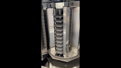
New rocking incubator for optimal brain organoid maturation
The CellXpress.ai system's new rocking incubator is a breakthrough for brain organoid culture. It supports dynamic motion within the incubator, allowing organoids to remain in constant movement throughout their development. The incubator can hold up to six racks, with a mix-and-match configuration that supports both static and rocking conditions. This means researchers can culture stem cells and brain organoids in the same incubator, with only the racks that require motion being rocked. Comparative studies have shown that organoids grown on a rocker are functionally and morphologically identical to those grown using an orbital shaker.
Previously, manually maintaining just 10 brain organoid plates, including daily feeding and imaging, required nearly 27 hours of hands-on time each week. Now, by automating the process, that same manual work is reduced to just a few hours.
Beyond time savings, the CellXpress.ai system brings scientific precision to brain organoid development. It automates feeding and imaging on a fixed schedule - including weekends - ensuring consistent treatment across samples and minimizing variability. The system also reduces cross-contamination risks, maintains optimal media conditions at 37°C, and captures full-well imaging with advanced feature analysis. Researchers can now track the entire cell journey over time, unlocking deeper insights while preserving culture integrity.