ON-DEMAND WEBINAR
High-content analysis and phenotypic characterization of 2D and 3D cellular models
As presented by Kayla Hill, PhD, Field Applications Scientist, Molecular Devices
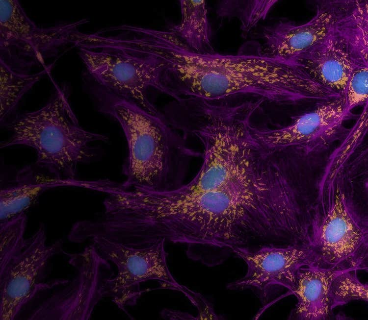
There is an increased need for expanding the variety and complexity of cell-based assays for biologic research and drug discovery. This has generated increasing interest in using three-dimensional (3D) cultures for assay development and phenotypic screening applicable for a range of cellular models including CNS, tumor micro-tissues, and organoids screens. Combining these 2D and 3D models with live-cell assays allow monitoring of cell responses in real time and provide important insights about compound treatment effects, biological complexity, and physiological relevance of assay results.
Key highlights:
- Use high-content analysis to analyze multiple parameters across high-throughput compound screens
- Overcome challenges in automating 3D organoid imaging with robust and flexible autofocusing methods and targeted imaging workflows
- Characterize compound effects on tumor spheroid phenotypes and volumetric changes in 3D cellular content with 3D image analysis algorithms
For more information on the complete line of Molecular Devices High Content Imaging Systems, please click here.
https://share.vidyard.com/watch/6XTZNzAxmwsrNM4hJP8RK1
Transcription of Question and Answer Session from Live Webinar
Q: What is the criteria threshold for the +/- cell nucleai staining?
A: Our Application Module looks at minimum and maximum width and intensity parameters.
Q: How do you achieve immunostatining within big 3D objects? Do you use reagents to clear organoids/sphreroids?
A: Clearing agents can help visualize deeper in the spheroid. Alternatively, culturing smaller spheroids will be beneficial if that is not an option.
Q: Is it possible to have different Z slicing settings for different X Y coordinates? Either different wells or different locations in the well
A: We can add an image-based focus to acquire images at different focal planes.
Q: Can you image 3D objects that are 4 - 5 mm in diameter?
A: We can image an object at 4-5 mm. The density and compactness of the object may affect how well we can visualize into the spheroid.
Q: What is the maximun Z step size for 10x objective?
A: There is no maximum step size at 10x. The limit is the working distance of the objective, which is 4mm.
Q: What is the limit in thickness you could image in an organoid? Specially in a non-cleared organoid?
A: We often image to an organoid depth of 100-150um to visualize the core. Other factors including compactness of the spheroid can have an impact.
Q: What are the NAs of the different lenses?
A: Our NAs range from 0.4 for our 2x Plan Apo to 1.2NA for our 60x Water Immersion lens.
Q: Is stitching of images at higher object magnification possible? Or you can only view the whole thing with lower objectives?
A: Yes, we can stitch or tile.
Q: Do you offer custom plates for your system or I could use Corning® ULA plates?
A: Our systems are plate agnostic. Corning ULA plates work well with the platform.
Q: Can the IXM XLS Fluidics that we already own go on the Micro Confocal?
A: Yes, we could use the existing fluidics on the XLS and put onto a new confocal system.
Q: Hello, what are your automation solutions for feeding plates into the micro confocal reader with a chamber?
A: We offer a Robo-EC option that is compatible with automation.
Q: Is it possible to use this imager for 'organ-on-chip' devices such as those offered by EmulateBio
A: Yes, we have imaged Emulate chips, which can be imaged directly from its designated holder.We have also imaged MIMETAS chips, as well as many other custom chip types.
Q: How do the transmitted light options compare to the ones offered in the micro XLS?Is it the same?
A: The Transmitted Light tower is the same as offered on the XLS. The LED option sits just below the plate door and enables compatibility with automation and fludics by eliminating the need for a physical tower on top of the instrument.
Q: Do you have a good way to do co-localizaton metrics?
A: We can address colocalization in a few different ways. One way is to score a target as positive for colocalization based on an intensity threshold. Alternatively, a translocation type module can be used to score based on correlation coefficient.
Q: If we upgrade our Micro XLS system to confocal, are the acqisition and analysis protocols easily importable into the Meta Xpress confocal software?
A: Yes, the acquisition modules and anlaysis protocols can be imported into a new version of MX. There just may be slight modifications to the settings.
Q: Can you add the fluid dispenser chamber to a IXMC?
A: Yes, the fluidics option can be added to an IXMC as an upgrade in the field.
Q: Hi, is the workflow to image a well at low res and then image areas with organoids at hi res a part of the regular software, or is that a customized solution you offer separately?
A: This is offered as part of the standard software package.
Q: I'm wondering how easy it is to set up the software to recognize cells within a matrix that is autofluorescent.
A: I would recommend using our confocal mode to remove haze from the matrix. We are able to detect cells quite well.
Q: At which point would you recommend having a laser vs LED as a light source?
A: A laser system is recommended if speed is a priority or if your signal is dim and you need a boost in S/N.
Q: In regards to upgrading from XLS to confocal, on the software side, is it only a software upgrade or add-on to existing software?Or it is a completely different software?
A: Depending on the version you are currently running, the software is very similar. We are on version 6 currently. If you were to upgrade to confocal you would get the latest software with win10 CPU.
Q: To reduce background from Matrix, are you aware of quenchers that could be added in your media?
A: I don't have any direct experience with using quenchers with a matrix, but we do offer some reagent kits wtih background quencher options.
Q: Can you easily swap an emission filter?
A: Yes emission filters can be swapped.
Q: Someone mentioned co-localization. Does that mean that traffiking to the membrane is detected?
A: The best way to analyze membrane trafficking is to identify the cell membrane with a membrane dye or whole cell marker. We can then create an analysis module to define the cell border and assess signal from the object of interest in the border
Q: Can you analyze a tagged protein?
A: We can certainly analyze expression of a tagged protein in a compartment of interest, in this case the membrane. All we need is a cell mask dye.
Q: Is the onboard LH field upgradable on a confocal?
A: Yes, it is.