
University of Leeds use ImageXpress Pico to investigate protein function
COMPANY/UNIVERSITY
University of Leeds
TEAM MEMBERS
Darren Tomlinson
Heather Martin
PRODUCTS USED
ImageXpress Pico Automated Cell Imaging System
The Challenge
The Tomlinson lab, in the School of Molecular and Cellular Biology at the University of Leeds, UK, investigates protein function at the molecular level. Darren Tomlinson’s research group is interested in using a novel molecular recognition tool, called an Affimer, to study protein function. An Affimer (also known as an Adhiron) is a scaffold protein that constrains two randomised loops for molecular recognition. They can be used as alternative reagents to antibodies for applications like diagnostics and used to probe the surface of target proteins to identify key domains involved in protein function. Their laboratory provides a service for screening against many types of molecules but their focus is using the Affimer scaffold to understand protein function at the molecular level.
To study these Affimers, the group needed an easy-to-use system that they could run small-scale screens on.
The Solution
The group chose the ImageXpress Pico as an affordable and user-friendly alternative to other high-content imaging systems.
ImageXpress Pico Automated Cell Imaging System
Products Used
The ImageXpress® Pico Automated Cell Imaging System is more than a digital microscope, combining high-resolution imaging with powerful analysis. Whether running fluorescence imaging or brightfield assays, the automated imager features a comprehensive portfolio of preconfigured protocols for cell-based assays to shorten the learning curve, so you can start running experiments quickly. With features such as Digital Confocal* 2D on-the-fly deconvolution, Live Preview, and multi-wavelength cell scoring, the ImageXpress Pico offers you the ability to advance your discoveries in a small, affordable imager..
The Results
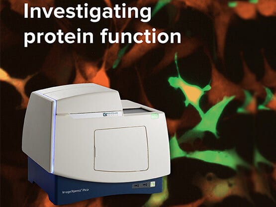
The group are primarily using the ImageXpress Pico for small-scale screens looking at the impact of Affimer proteins targeting a variety of intracellular proteins on the signaling pathways downstream of the targeted proteins. An example can be seen in figure 1, which shows mouse embryonic fibroblasts transfected with GFP-tagged Affimers (green) stained for phosphorylated ERK (orange). It demonstrates that the protein targeting Affimer reduces EGF-stimulated pERK expression.
The group also use the ImageXpress Pico for time-course experiments, where the temperature control feature comes in particularly useful.
They are now imaging approximately 2-3 plates per day, depending on the assay as they tend to use 3-4 fluorophores/plate.

Figure 1: Mouse embryonic fibroblasts transfected with GFP-tagged Affimers (green) and stained for phosphorylated ERK (orange). Arrows highlight Affimer expressing cells where a reduction in pERK staining is clearly observed. Images acquired on the ImageXpress Pico at 20x magnification.