
Application Note
Highly sensitive multiplex TR-FRET assay on the SpectraMax iD5e Multi-Mode Microplate Reader
- Save valuable samples by measuring two analytes in the same well
- Reduce time spent on sample and plate preparation
- Reduce costs by using fewer plates and consumables, along with a cost-efficient kit
- Streamline your assays with automated data acquisition and analysis using SoftMax® Pro Software
Stanimira Valeva, PhD | Field Applications Scientist | Molecular Devices
Caroline Cardonnel, PhD | Application Scientist Supervisor | Molecular Devices
Susana Brun, PhD | Biology R&D Manager | Poly-Dtech
Mohamadou Sy, PhD | Chemistry R&D Manager | Poly-Dtech
Joan Goetz, PhD | CEO | Poly-Dtech
Introduction
In this application note, we demonstrate how the SpectraMax® iD5e Multi-Mode Microplate Reader can be used to perform a robust, no-wash immunoassay with detection of two targets in the same well using Multi-Dtech™ technology from Poly-Dtech.
Time-Resolved Förster Resonance Energy Transfer (TR-FRET) is a cutting-edge immunoassay technique widely used for the detection and quantification of biomolecules. This homogeneous method is fast, wash-free, reduces background fluorescence, and is cost-effective. Bright-Dtech™ technology incorporates high-brightness nanoparticles and fluorophores, improving the limits of detection and sensitivity compared to traditional TR-FRET methods.
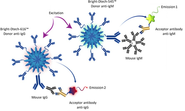
Figure 1. Multiplex TR-FRET technology using Bright-Dtech nanoparticles
In the Multi-Dtech TR-FRET method, two pairs of antibodies are employed to specifically detect mouse IgM and IgG. The donor antibodies are conjugated to lanthanide-based fluorescent nanoparticles, while the acceptor antibodies are labelled with fluorophores emitting in the green (520 nm) or red (665 nm) spectral region. When the targets are recognized by the antibody pairs, energy is transferred from the donor to the acceptor, generating two distinct signals corresponding to each analyte (Figure 1). These signals are simultaneously measured using time-resolved fluorescence, yielding highly specific and quantitative results due to reduced background fluorescence and an improved signal-to-noise ratio compared with conventional fluorescence detection (Figure 2).
The Mouse IgG/IgM Assay Kit enables the simultaneous quantitative detection of mouse IgG and IgM in serum using TR-FRET methodology. This kit incorporates Bright-Dtech technology, which enhances detection through its exceptional specificity and sensitivity. The assay dynamic range spans from 1 to 1000 ng/mL.
Materials
- Multi-Dtech Multiplex TR-FRET Assay Kit Mouse IgM/IgG (Poly-Dtech P/N FRETmlgG/IgM)
- Nunc™ 384 Well ShallowWell Standard Height Black (Thermo Scientific P/N 264705) included with the Multi-Dtech Multiplex TR-FRET Assay Kit
- SpectraMax iD5e Multi-Mode Microplate Reader*, with
- Enhanced TRF Module
- (Molecular Devices P/N 0200-7030)
- Time Resolved Filter 340nm BW 70nm (Molecular Devices P/N 6590-0080)
- Time Resolved Filter 616nm BW 10nm (Molecular Devices P/N 6590-0118)
- Time Resolved Filter 665nm BW 10nm (Molecular Devices P/N 6590-0121)
- Time Resolved Filter 520nm BW 15nm (Molecular Devices P/N 6590-0098)
- Fluorescence, Luminescence, and TRF Filter 535nm BW 25nm (P/N 6590-0099)
- SoftMax Pro data acquisitions and analysis software (Molecular Devices)
*Equivalent results are generated with the SpectraMax iD5 reader
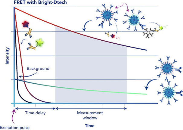
Figure 2. Principle of time-resolved signal measurement using Bright-Dtech nanoparticles. Time-resolved fluorescence (TRF) reduces background by using a lanthanide fluorophore, such as europium or terbium, which emits long-lived fluorescence. This fluorescence lasts for milliseconds; therefore, excitation by a pulsed light source (e.g., flash lamp), followed by a delay and then a signal measurement (‘counting window’), allows short-lived background fluorescence (lasting only nanoseconds) to decay before the signal is recorded.
Methods
The Multiplex TR-FRET mouse IgM/IgG assay was performed by Poly-Dtech as described in the kit data sheet protocol, available on the manufacturer’s website. IgM and IgG standards were prepared at final concentrations from 0 to 1000 ng/mL using 2-fold serial dilutions with Poly-Dtech TRF assay buffer. The standards were dispensed at their final concentrations in duplicate at 10 µL per well, followed by 5 μL of the Donor/Acceptor mix solution.
The plate was covered with adhesive foil and incubated for 1.5 hours at room temperature. Time-resolved fluorescence was measured using the SpectraMax iD5e reader (see Table 1 for instrument settings). Both Microplate Optimization and Read Height adjustment were performed using the SpectraMax iD5e reader to ensure optimal assay sensitivity and dynamic range.
Data analysis
The TR-FRET ratio was calculated for each well using the following formula:
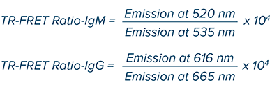
This ratiometric measurement reduces well-to-well variability and minimizes potential signal interference. The intensity of the resulting signal is directly proportional to the concentration of the antigen present in the sample.
Standard curves for IgM and IgG were generated by plotting the TR-FRET ratio on the Y-axis and the log10 of Ig concentration in the X-axis using a 4-parameter logistic (4PL) curve fit.
The limit of detection (LOD) was determined by adding three (IgG) or four (IgM) standard deviations to the mean value of the blank (zero) replicates and calculating the corresponding concentration.
The limit of quantification (LOQ) was determined by adding ten standard deviations to the mean value of the blank (zero) replicates and calculating the corresponding concentration.
All data were generated and analyzed using SoftMax Pro Software.
Results
The data were analyzed as described above and plotted in SoftMax Pro software using a 4-parameter logistic (4PL) curve fit for IgM and IgG (Figure 3). Both calibration curves showed excellent linearity, with R2 = 0.999 and %CV ≤ 5%. The EC50 values and LOD are presented in Table 2. The assay window for IgM was larger; however, both targets were detected with excellent sensitivity and low signal variability.
Select ‘Use Filter’ for both Ex, Em.
IgM detection:
Excitation: 340 nm
Emission 1: 535 nm
Emission 2: 520 nm
IgG detection:
Excitation: 340 nm
Emission 1: 616 nm
Emission 2: 665 nm
Excitation time: 0.05 ms
Measurement delay: 0.03 ms
Integration time: 0.4 ms
Read height [optimize]
Table 1. Optimized instrument settings for the Poly-Dtech Multiplex TR-FRET assay on the SpectraMax iD5e reader.
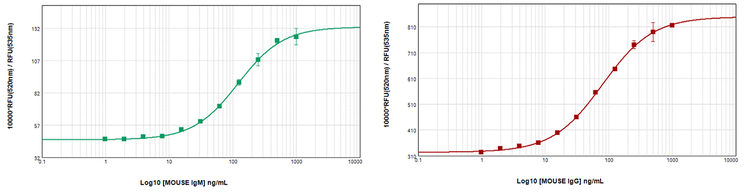
Figure 3. Mouse IgM (green) and IgG (red) calibration curves in a multiplex assay. The curves were generated and measured in the same wells. The Y-axis reflects TR-FRET ratio as calculated in the Methods section. Both curves showed R2 = 0.999 and CV% ≤ 5% for all replicates. Both curves showed parameter independencies of at least 0.9, demonstrating the goodness of the fit.
Table 2. Assay parameters for IgM and IgG Poly-Dtech multiplex assay on SpectraMax iD5e. The assay parameters validated by Poly-Dtech are as follows: EC50: 110 ng/mL for IgM and 79 ng/mL for IgG; LOD: 2 ng/mL (IgM) and 1.7 ng/mL (IgG).
Conclusion
The enhanced sensitivity of Bright-Dtech™ nanoparticles enables the detection of low analyte concentrations with high accuracy. By combining green (terbium) and red (europium) detection, the Multiplex TR-FRET assay developed by Poly-Dtech saves time and precious samples while maintaining high assay quality and sensitivity, as demonstrated by the excellent curve fit and low LOD obtained using the SpectraMax iD5e reader.
Optimized data acquisition and automated data analysis workflows can be saved as protocols in SoftMax Pro software for repeated use, ensuring consistent and efficient results.
References
- https://poly-dtech.com/product/multi-dtech-mouse-igg-igm/
- Joan Goetz, Aline Nonat, Abdoulaye Diallo, Mohamadou Sy, Ildan Sera, Alexandre Lecointre, Christophe Lefevre, Chi Fai Chan, Ka-Leung Wong, Loïc J Charbonnière, (2016). Ultra-bright lanthanide nanoparticles. Chempluschem. 2016 Jun;81(6):497. doi: 10.1002/ cplu.201600117
- Cyrille Charpentier, Vjona Cifliku, Joan Goetz, Aline Nonat, Clémence Cheignon, Marcelina Cardoso Dos Santos, Laura Francés-Soriano, Ka-Leung Wong, Loïc J Charbonnière, Niko Hildebrandt (2020). Ultrabright Terbium Nanoparticles for FRET Biosensing and In Situ Imaging of Epidermal Growth Factor Receptors. Chemistry. 2020 Nov 17;26(64):14602-14611. doi: 10.1002/chem.202002007