Application Modules for MetaXpress and MetaXpress PowerCore Software
Application modules for the MetaXpress® and MetaXpress® PowerCore™ software allows users to easily automate common
analysis tasks. The following modules are now available:

Application modules feature:
- Intuitive dialog boxes
- Adaptive Background Correction - Improved segmentation through adaptation to local content
- Assay can be run across entire data set
- Field and cell-by-cell data logging
- Compatibility with MetaXpress PowerCore high throughput image analysis
- Customization through MetaMorph® software journals (not compatible with MetaXpress PowerCore software)
Typical applications include:
- Endothelial tube formation: Angiogenesis tube formation
- Promotion or inhibition of blood vessel formation: Angiogenesis tube formation
- Cell viability: Live/dead, Cell cycle, Multi Wavelength Cell Scoring
- Cell cycle: Mitotic Index, Cell cycle
- Membrane potential: Cell health
- Cytotoxicity and apoptosis: Live/dead
- Cell proliferation: Count nuclei, Cell Proliferation HT, Live/dead
- Cell migration: Count nuclei, Cell Proliferation HT
- Cell counting: Count nuclei, Cell Proliferation HT
- Determination of cell health and proliferation in populations of mono-, bi- and multi-nucleated cells: Micronuclei
- Micronuclei and genotoxicity analysis: Micronuclei
- Kinase activation: Cell scoring
- Fatty acid uptake: Cell scoring, Multi Wavelength Cell Scoring
- Mitosis: Mitotic Index
- Adipogenesis: Cell scoring, Multi Wavelength Cell Scoring
- Examining transfection efficiencies: Cell scoring, Multi Wavelength Cell Scoring
- Studying intracellular structures: Transfluor, Transfluor HT, Granularity
- Receptor internalization: Transfluor, Transfluor HT, Granularity
- Punctate staining: Transfluor, Transfluor HT, Granularity
- Clustering target molecules: Transfluor, Transfluor HT, Granularity
- Process extension: Neurite outgrowth
- Neurodegenerative or neuroregenerative disease research: Neurite outgrowth
- Cell differentiation (Stem cell research): Neurite outgrowth
- Protein movement: Translocation, Nuclear Translocation HT, Multi Wavelength Translocation
- Cell pathway analysis: Translocation, Nuclear Translocation HT
- Protein phosphorylation: Cell scoring, Multi Wavelength Cell Scoring
- Mitochondrial localization: Translocation
- Detect monopolar spindles: Monopole detection
Micronuclei application module
- Classify Micro- and Multi-nucleated cells
- Additional markers for analysis of cytotoxicity
- More than 100 outputs
- Fastest "acquisition through analysis" workflow with on-the-fly cell counting and MetaXpress® PowerCore™ high-throughput image analysis
Angiogenesis tube formation application module
- Better quantitation by creating a single in-focus composite image from multiple Z-series images
- Multi-parameter analysis measurements include tube length, number of branch points, number of nodes and more
|
Figure 1. Inhibition of Human Mammary Epithelial Cells (HMEC-1)
tube formation by suramin. BD BioCoat™ Angiogenesis System.


Data courtesy of BD Biosciences.
|
Cell cycle application module
- Quantitation of cell cycle stages
- Optional apoptosis and mitotis-specific stains
|
Figure 2. Cell cycle classification can be performed with a single or multi wavelength assay. Left: CHO-K1 cells stained with Hoechst 33342 were imaged with a 20x objective on the Discovery-1 system using MetaXpress software. Right: The Cell Cycle module identifies cell cycle phases: G0/G1 (dark blue), S (light blue), G2 (green), Early M (orange) and Late M (red).
|
Cell health application module
- Analysis of up to three fluorescent probes for cell-based assays of apoptosis and necrosis
- Multi-parameter analysis measurements include counts and percentages of viable, early and late apoptotic and necrotic cells and more
|
Figure 3. Adherent Chinese Hamster Ovary (CHO-K1) cells were incubated with various concentrations of staurosporine
for 6-12 hours. Top left: control, top right: 0.1 µM staurosporine, bottom left: 3 µM staurosporine,
bottom right: measured (Green: viable, blue: early apoptotic, purple: late apoptotic, red: necrotic).
|
Cell scoring application module
- Identification of two sub-population of cells
- Ideal for counting and logging measurements of cells in two-wavelength experiments
|
Figure 4. The module can be used as a generic segmentation tool
to identify two different stains (top) or to obtain cell details (bottom).
|
Count nuclei application module
- Automatically counts nuclei and captures intensity measurements
- Accurate segmentation of touching cells
|
Figure 5. Left: Image has uneven illumination and touching cells. Right: Count Nuclei identifies touching
cells as separate objects (as indicated by the red arrow). Adaptive Background Correction™ compensates for
the variable background intensity even when background intensity in one area of the image is greater than
nuclei intensity in another area.
|
Live/dead application module
- Quantitation of live and dead cells
- Multi-parameter analysis measurements include quantitation and percentages of live and dead cells, per-wavelength measurements and more
|
Figure 6. Adherent Chinese Hamster Ovary (CHO-K1) cells were incubated with varying concentrations of
DMSO for 6-12 hours prior to staining. Cells were labeled with H33342 and PI diluted in PBS for 30-60 minutes @RT
before image acquisition. Top left: control, top right: 20% DMSO, bottom: measured (red: dead, green: live).
|
Mitotic index application module
- Quantitation and percentage of mitotic and interphase cells
- Measurements include count, percentage and wavelength-specific intensities of mitotic and non-mitotic nuclei, and more
|
Figure 7. Left: CHO-K1 cells treated with Nocadazole for 18 hours before staining with anti-phospho-Histone H3
(Ser28) and acquired by Discovery-1. Right: mitotic (green) and interphase (red) cells.
|
Monopole detection application module
- Quantitation of mitotic cells with monopolar or bipolar spindles
- Measurements include count and percentage of monopoles, bipoles and interphase cells, DNA structures area, cell classification and more
|
Figure 8. 3T3-L1 mouse fibroblast cells treated with monastrol and stained with mouse anti-beta tubulin
primary antibody detected with a FITC conjugated goat anti-mouse secondary antibody. Nuclei are stained with
Hoeschst 33342. Left: monastrol, right: segmented image shows interphase cells (red), bipolar spindles (blue)
and monopole (green).
|
Neurite outgrowth application module
- Multi-parameter analysis measurements include total neurite outgrowth, total branches and cell bodies, straightness and more
- Works with or without nuclear stain
|
Figure 9. Top: DF cells. Bottom: DF cells skeletonized.

Data courtesy of Rinat Neuroscience Corporation.
|
Transfluor application module
- Designed to facilitate the analysis of receptor internalization specifically for the Transfluor assay
- Intensity measurements and quantitation of pits, vesicles and nuclei
- Choice of six granularity indices
|
Figure 10. Human osteosarcoma cells (U2OS) incubated overnight for 40-45 minutes (vesicle formers) or
30 minutes (pit formers). Varying concentrations of isoproterenol were added to one set of wells. Top: acquisition,
bottom: Pits (white), vesicles (red) and nuclei (green) can be simultaneously segmented. Grains are accurately
identified even in cells with high background (red arrow).
|
Translocation application module
- Quantitation of correlation between probes and compartments
- Two modules to fit your needs: the Translocation module, ideal for images that need a minimum amount of adjustments, and the Translocation-Enhanced module, for additional flexibility
- Measurements include compartments, mean compartment area, correlation coefficient, intensity, number and percentage of positives, and more
|
Figure 11. Mitochondrial translocation. HeLa cells treated with Staurosporine and stained with MitoTracker
(red), DAPI (blue) and anti-Cytochrome C (green).
|
Ordering Information
|
Product Name
|
Part Number
|
|
MetaXpress Offline Analysis Station with MDCStore Integrated Database Functionality
|
METAX OFFLINE
|
|
Angiogenesis Tube Formation
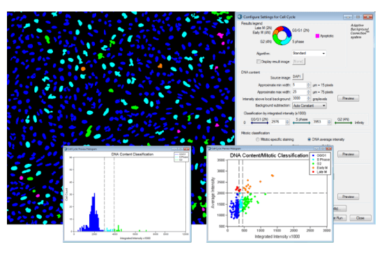
 Application Module |
9500-0016
|
|
Cell Cycle Application Module |
9500-0040
|
|
Cell Health Application Module |
9500-0035
|
|
Cell Proliferation HT Application Module
|
9500-0042
|
|
Cell Scoring Application Module |
9500-0037
|
|
Count Nuclei Application Module |
9500-0017
|
|
Granularity Application Module |
9500-0032
|
|
Live/Dead Application Module |
9500-0034
|
|
Micronuclei Application Module |
9500-0045
|
|
Mitotic Index Application Module |
9500-0036
|
|
Monopole Detection Application Module
|
9500-0039
|
|
Multi Wavelength Cell Scoring Application Module
|
9500-0038
|
| Multi Wavelength Translocation Application Module |
9500-0044 |
|
Neurite Outgrowth Application Module |
9500-0015
|
|
Nuclear Translocation HT Application Module
|
9500-0041
|
|
Transfluor Application Module |
9500-0033
|
|
Transfluor HT Application Module
|
9500-0043
|
|
Translocation Application Modules |
9500-0014
|
To place an order, call your local Molecular Devices Office or
Molecular Devices Distributor.
|